 뒤로
Cardiovascular System: Medical Terminology Study Guide
스터디 가이드 - 스마트 노트
 자료에 맞춘 맞춤형 노트, 핵심 정의, 예시, 맥락을 확장해 제공합니다.
Cardiovascular System Overview
Introduction to the Cardiovascular System
The cardiovascular system is responsible for maintaining blood circulation throughout the body. It consists of the heart, blood vessels, and blood, and performs several essential functions:
Nutrient Transport: Delivers oxygen and essential nutrients, enzymes, and hormones to tissues.
Protection: Helps regulate body temperature and defends against pathogen invasion.
Waste Removal: Transports carbon dioxide to the lungs and other waste products to the liver and kidneys for excretion.

Example: The cardiovascular system does not deliver digestive enzymes like salivary amylase to the stomach lumen.
Cardiovascular System Divisions
Pulmonary and Systemic Circuits
The cardiovascular system is divided into two main circuits:
Pulmonary Circuit: Carries oxygen-poor blood from the heart to the lungs for gas exchange and returns oxygen-rich blood to the heart.
Systemic Circuit: Distributes oxygen-rich blood from the heart to the rest of the body and returns oxygen-poor blood back to the heart.
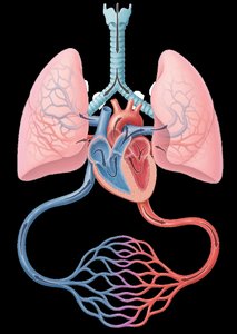
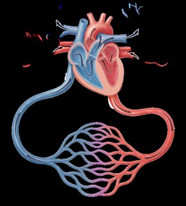
Example: The pulmonary circuit transports carbon dioxide-rich blood from the heart to the lungs for gas exchange.
Blood Vessels
Types and Functions of Blood Vessels
Blood vessels are tube-like structures forming a network to transport blood throughout the body. They originate in the heart, branch throughout the body, and return blood to the heart. There are five main types:
Arteries: Carry blood away from the heart.
Arterioles: Small arteries that branch from larger arteries.
Capillaries: Smallest blood vessels, site of exchange between blood and tissues.
Venules: Small veins that collect blood from capillaries.
Veins: Carry blood toward the heart.
Example: Capillaries nourish living cells in hair roots (papilla).
Practice: The branching of arteries into smaller vessels ensures that blood reaches all parts of the body.
Heart Structure
Anatomy of the Heart
The heart is the central organ of the cardiovascular system, primarily composed of cardiac muscle. It has four chambers and three layers:
Atria: Two upper chambers that receive blood.
Ventricles: Two lower chambers that pump blood out.
Endocardium: Lines heart chambers, reduces inner friction.
Myocardium: Thick muscular layer responsible for contraction.
Pericardium: Double-walled fluid-filled sac, reduces outer friction.
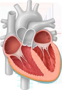
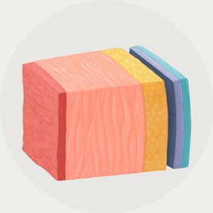
Example: The pericardium protects the myocardium, which is lined by the endocardium. The atria receive blood, and the ventricles pump blood.
Heart Valves
Types and Functions of Heart Valves
Heart valves are located at the entrances and exits of the heart chambers, controlling the direction of blood flow:
Atrioventricular Valves: Allow blood received in the atria to enter the ventricles (Tricuspid and Bicuspid/Mitral valves).
Semilunar Valves: Allow blood to flow out of the ventricles (Aortic and Pulmonary valves).
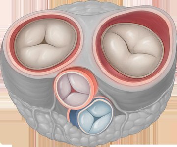
Example: The tricuspid valve is named for its three cusps. Semilunar valves allow blood to exit the ventricles.
Practice: Heart valves ensure one-way blood flow; the atria receive blood, and the ventricles pump it out.
Major Arteries and Veins
Major Arteries
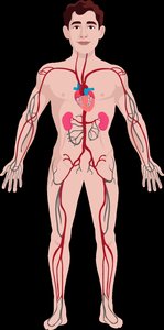
Arteries are blood vessels that carry blood away from the heart. Major arteries include:
Aorta: Main artery leaving the heart.
Carotid: Supplies the head and neck.
Brachial, Radial, Ulnar: Supply the arms.
Renal: Supplies the kidneys.
Femoral, Popliteal, Tibial, Peroneal: Supply the legs.
Major Veins
Veins return blood to the heart. Major veins include:
Jugular: Drains the head and neck.
Subclavian, Axillary, Brachial, Radial, Ulnar: Drain the arms.
Superior and Inferior Vena Cava: Largest veins returning blood to the heart.
Femoral, Popliteal, Tibial, Great Saphenous: Drain the legs.
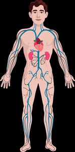

Example: The great saphenous vein is a superficial vein on the medial side of the leg.
Practice: The azygos vein is an unpaired blood vessel.
Medical Terminology: Word Building
Combining Forms for Cardiovascular Structures
Combining forms are used to build medical terms related to the cardiovascular system:
Combining Form | Definition |
|---|---|
coron/o | heart |
vas/o, vascul/o, angi/o | vessel |
ven/o, phleb/o | vein |
sept/o | wall, partition |
sphygm/o | pulse |
steth/o | chest |
corpor/o | body |

Example: A specialist trained to withdraw blood from veins is a phlebotomist.
Practice: Extracorporeal circulation refers to blood being circulated and oxygenated outside the body.
Combining Forms for Pathology
Combining Form | Definition |
|---|---|
aneurysm/o | widened blood vessel |
varic/o | varicose vein |
man/o | pressure |
sten/o | narrow |
ather/o | fatty substance |
infarct/o | necrosis of tissue in an area |
occlus/o | to close up |
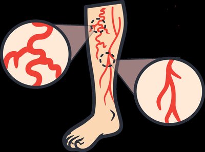


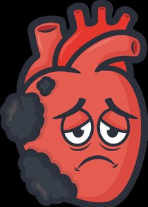

Example: Myocardial infarction is tissue death due to blocked blood flow to the heart muscle.
Practice: Atherosclerosis is the buildup of fatty deposits in arteries, causing them to harden and narrow.
Suffixes Used in Cardiovascular Terms
Suffix | Definition |
|---|---|
-cardia | heart condition |
-pressor | to press down |
-ectasis | dilatation |

Example: Coronary artery ectasis is dilation of a blood vessel. A vasopressor is a medication that constricts blood vessels to increase blood pressure.
Practice: Ventricular tachycardia is a fast heart rhythm originating in the lower chambers.
Cardiovascular Pathology
Clinical Disorders
Medical Term | Definition |
|---|---|
Congestive Heart Failure | Reduced outflow from one side of the heart due to myocardial damage. |
Peripheral Vascular Disease | Plaque buildup in blood vessels outside the heart, often affecting the legs. |
Patent Ductus Arteriosus (PDA) | Congenital defect where a blood vessel fails to close after birth. |
Example: Raynaud Phenomenon is characterized by spasms of arterioles in the fingers, sometimes leading to tissue damage.
Practice: Symptoms of heart failure include persistent cough with mucus and shortness of breath, but not excessive thirst or weight loss.
Cardiovascular Diagnostic and Laboratory Terms
Diagnostic and Surgical Procedures
Medical Term | Definition |
|---|---|
Thallium Stress Test (TST) | Radiological cardiac scan measuring blood flow during exercise and rest. |
Holter Monitor | Portable device that detects heart rhythm abnormalities. |
Automated External Defibrillator (AED) | Portable device delivering an electrical shock to restore heart rhythm. |
Coronary Artery Bypass | Surgical procedure creating a new pathway for blood flow using a vessel. |
Example: Thallium-201 is administered intravenously for a stress test.
Practice: The primary function of an AED is to restore a regular heart rhythm.
Cardiovascular Pharmacology
Drug Classes and Their Effects
Drug Class | Description | Function |
|---|---|---|
ACE Inhibitors | Expand blood vessels | Lower blood pressure, reduce fluid retention, protect kidneys |
Calcium Channel Blockers (CCB) | Block inward movement of calcium ions | Reduce force of contractions and heart rate |
Digitalis Drugs | Increase force of heart contractions | Strengthen heart muscle |
Example: Calcium channel blockers weaken cardiac muscle contraction by blocking calcium ions.
Practice: ACE inhibitors are vasodilators.
Additional info: All content is expanded and structured for clarity, with relevant images included only where they directly reinforce the educational content.
