 뒤로
뒤로Chapter 12: The Special Senses – Medical Terminology Study Guide
스터디 가이드 - 스마트 노트
 자료에 맞춘 맞춤형 노트, 핵심 정의, 예시, 맥락을 확장해 제공합니다.
The Special Senses
Overview of General vs. Special Senses
The human body detects environmental changes through specialized sensory receptors. These are classified as general senses and special senses, each with distinct anatomical locations and functions.
General Sense Receptors: Distributed throughout the body, including skin, muscles, joints, and visceral organs. Responsible for detecting touch, pain, temperature, and proprioception.
Special Sense Receptors: Located in dedicated organs of the head. The five special senses are olfaction (smell), gustation (taste), vision, hearing, and equilibrium. Signals are relayed to the brain via cranial nerves.
Receptor Modalities
Sensory receptors are classified by the type of stimulus they detect:
Chemoreceptors: Detect chemicals in bodily fluids or the environment (e.g., taste, smell, blood pH).
Thermoreceptors: Detect temperature changes.
Photoreceptors: Detect light intensity, color, and movement; found only in the retina.
Mechanoreceptors: Detect pressure and mechanical forces.
Nociceptors: Detect painful stimuli.
Proprioceptors: Detect balance and posture; located in tendons and muscles.
Olfaction (Smell)
Anatomy and Function of Olfactory Structures
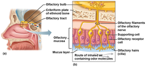
Olfaction is the sense of smell, mediated by specialized structures in the nasal cavity. The olfactory membrane contains receptor cells that bind odor molecules, allowing the perception of thousands of distinct odors.
Olfactory Membrane: Located on the roof of the nasal cavity.
Olfactory Receptor Cells: Sensory neurons with olfactory hairs (cilia) that act as chemoreceptors.
Olfactory Bulb and Tract: Relay signals to the brain.
Supporting Cells: Provide structural and metabolic support.
Mucus Layer: Traps and dissolves odor molecules for detection.
Gustation (Taste)
Anatomy and Function of Gustatory Structures
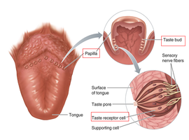
Gustation is the sense of taste, primarily mediated by taste buds located on the tongue and oral cavity. Each taste bud contains multiple receptor cells capable of detecting different taste molecules.
Tongue Papillae: Bumps on the tongue surface housing taste buds.
Taste Buds: Onion-shaped structures containing gustatory cells.
Taste Receptor Cells: Detect taste molecules and relay signals to sensory nerve fibers.
Supporting Cells: Provide structural support within the taste bud.
The Five Basic Tastes
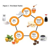
Humans can detect five primary tastes, each associated with specific molecules:
Sweet: Sugars and sweeteners
Salty: Metal ions (e.g., sodium, potassium)
Sour: Acids
Bitter: Bases and certain alkaloids
Umami: Amino acids, "meaty" taste
Interaction of Smell and Taste
Flavor perception is a combination of taste, smell, and texture. While taste buds detect five basic flavors, olfactory receptors contribute to the nuanced identification of food flavors. Blocking olfactory receptors impairs flavor detection.
Example: Identifying the specific flavor of a jellybean relies on olfactory input, not just taste.
Vision
Anatomy of the Eye
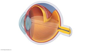
The eye is a complex organ with three main layers, each contributing to vision. Light is focused onto the retina, where photoreceptors convert it into neural signals.
Fibrous Layer: Includes the cornea (clear, refracts light) and sclera (white, tough connective tissue).
Vascular Layer: Includes the ciliary body (controls lens size), iris (adjusts pupil diameter), choroid (absorbs excess light), and lens (focuses light).
Neural Layer: Includes the retina (houses photoreceptors), optic nerve (relays signals), and macula lutea (area of high cone density for clear vision).
Visual Pathway and Eye Reflexes
Light travels through the cornea, pupil, lens, vitreous humor, and retina before reaching the optic nerve and brain. The eye adapts to varying light conditions and focuses on objects at different distances.
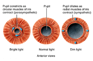
Pupillary Reflex: Iris muscles constrict or dilate the pupil to regulate light entry.
Visual Accommodation: Lens changes shape to focus on near or far objects.
Fluids of the Eye
The eye contains two main fluids that help maintain its shape and optical properties:
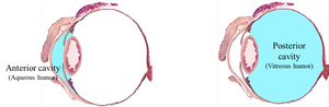
Vitreous Humor: Gelatinous fluid in the posterior cavity (behind the lens).
Aqueous Humor: Watery fluid in the anterior cavity (in front of the lens).
Glaucoma: Disease caused by elevated intraocular pressure, potentially damaging vision.
Photoreceptors: Rods and Cones
The retina contains two main types of photoreceptors:
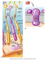
Rods: Function in dim light, detect grayscale, produce blurry images.
Cones: Function in bright light, detect color, produce high-resolution images. Concentrated in the macula lutea and fovea centralis.
Color Vision and Colorblindness
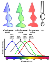
Three types of cones allow color vision, each sensitive to a specific wavelength:
Blue Cones: Absorb blue wavelengths (~427 nm)
Green Cones: Absorb green wavelengths (~533 nm)
Red Cones: Absorb red wavelengths (~564 nm)
Red-Green Colorblindness: Genetic disorder affecting green and/or red cones.
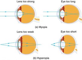
Visual Impairments
Proper focusing of light onto the fovea is essential for clear vision. Common impairments include:
Myopia (Near-sightedness): Eyeball too thick or lens too strong; distant objects appear blurry.
Hyperopia (Far-sightedness): Eyeball too short or lens too weak; close objects appear blurry.
Correction: Glasses, contacts, or laser surgery adjust light focus.
Hearing
Anatomy of the Ear
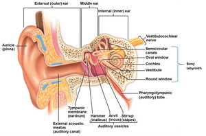
The ear is divided into three main regions, each with specialized structures for sound detection and transmission.
Outer Ear: Auricle (ear lobe) and external acoustic meatus (ear canal).
Middle Ear: Tympanum (ear drum), auditory ossicles (malleus, incus, stapes), pharyngotympanic tube (equalizes pressure).
Inner Ear: Bony labyrinth, cochlea (contains hair cells for sound detection), vestibulocochlear nerve (CNVIII).
Hearing Pathway
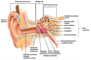
Sound waves are transmitted through the ear structures to the brain:
Sound waves strike the tympanic membrane.
Vibrations are conducted by the malleus, incus, and stapes.
Stapes transmits vibrations to the oval window.
Hair cells in the cochlea detect sound frequencies.
Signals are relayed via the vestibulocochlear nerve to the temporal lobe.
Equilibrium
Anatomy and Function of Equilibrium Structures
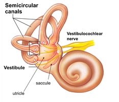
Equilibrium is the sense of balance, mediated by fluid-filled chambers in the inner ear and proprioceptors in muscles.
Semicircular Canals and Vestibule: Contain receptors that detect changes in head position and movement.
Proprioceptors: Located in muscles, contribute to balance and posture.
Photoreceptors: In the eyes, also play a role in equilibrium.
Summary Table: Special Senses and Their Main Structures
Sense | Main Organ | Receptor Type | Key Structure |
|---|---|---|---|
Olfaction | Nasal cavity | Chemoreceptor | Olfactory membrane, olfactory receptor cells |
Gustation | Tongue | Chemoreceptor | Taste buds, gustatory cells |
Vision | Eye | Photoreceptor | Retina, rods and cones |
Hearing | Ear | Mechanoreceptor | Cochlea, hair cells |
Equilibrium | Inner ear | Mechanoreceptor, proprioceptor | Semicircular canals, vestibule |
Practice Questions
Which structure controls the size of the lens? D) ciliary body
What is the correct light wave pathway to the brain? A) cornea → pupil → lens → vitreous humor → retina → optic nerve
Sense of equilibrium requires __________? D) all of these (cochlea, vestibule, semicircular canals)
