 뒤로
뒤로Neurological System: Medical Terminology and Clinical Concepts
스터디 가이드 - 스마트 노트
 자료에 맞춘 맞춤형 노트, 핵심 정의, 예시, 맥락을 확장해 제공합니다.
Brain Cells and Neuroglia
Types of Brain Cells
The brain is composed of two main types of cells: neurons and glial cells. Neurons are the primary messengers of the nervous system, responsible for transmitting information. Glial cells provide support, protection, and nutrition to neurons.
Neurons: Specialized for receiving, conducting, and transmitting electrical signals.
Glial Cells: Include oligodendrocytes, microglia, and astrocytes, each with distinct functions.
Example: Oligodendrocytes form the myelin sheath around axons, speeding up signal transmission. 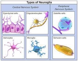
Parts of a Neuron
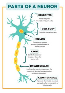
Neurons have specialized structures for their function:
Dendrites: Receive signals from other neurons.
Cell Body: Contains the nucleus and genetic material.
Axon: Conducts electrical impulses away from the cell body.
Myelin Sheath: Insulates the axon, increasing impulse speed.
Axon Terminal: Transmits signals to other cells.
Central Nervous System (CNS)
Regions of the Brain
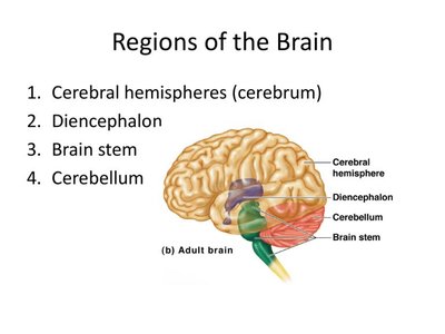
The CNS consists of the brain and spinal cord. The brain is divided into four main regions:
Cerebrum: Largest part, responsible for higher functions.
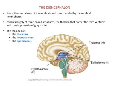
Diencephalon: Contains thalamus, hypothalamus, and epithalamus.
Brainstem: Controls vital functions.
Cerebellum: Coordinates movement and balance.
Cerebrum and Its Lobes
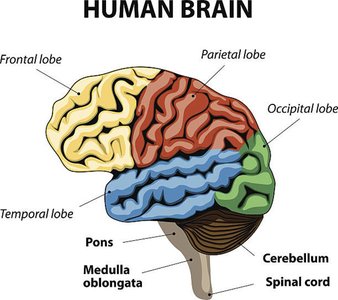
The cerebrum is divided into right and left hemispheres and four lobes:
Frontal Lobe: Motor control, decision-making, personality.
Parietal Lobe: Processes sensory information.
Temporal Lobe: Auditory processing, speech comprehension.
Occipital Lobe: Visual processing.
Diencephalon
The diencephalon is located centrally within the cerebrum and includes:
Thalamus: Sorts and directs sensory input.
Hypothalamus: Regulates homeostasis, stress response, and circadian rhythms.
Pituitary Gland: Releases hormones for homeostasis.
Epithalamus: Involved in sleep-wake cycles and emotional processes.
Pineal Gland: Secretes melatonin.
Cerebellum
The cerebellum, or "little brain," is responsible for coordination, balance, postural stability, and fine motor movement. 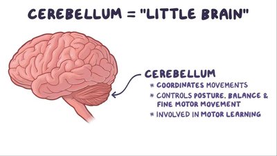
Brainstem
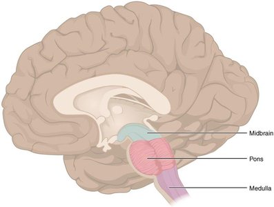
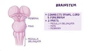
The brainstem consists of the midbrain, pons, and medulla oblongata. It controls vital functions such as heart rate, blood pressure, respiration, and reflexes.
Midbrain: Auditory and visual functions.
Pons: Controls respiration and sleep-wake cycle.
Medulla Oblongata: Regulates heart rate, blood pressure, and involuntary reflexes.
Meninges
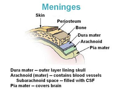
The meninges are protective connective tissue layers covering the brain and spinal cord.
Dura Mater: Tough, outer layer.
Arachnoid Mater: Middle, delicate layer.
Pia Mater: Inner layer, rich in blood vessels.
Ventricles & Cerebrospinal Fluid (CSF)
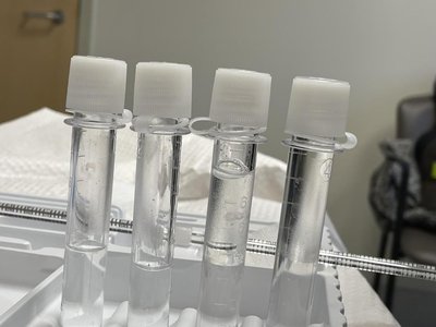
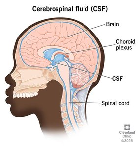
The brain contains four ventricles that produce and circulate CSF, which cushions, nourishes, and protects the CNS.
CSF: Clear fluid produced in the choroid plexus, circulates through ventricles and subarachnoid space.
Functions: Protection, nutrient delivery, waste removal, and regulation of intracranial pressure.
Spinal Cord & Spine
The spinal cord is a cylindrical structure within the spinal column, transmitting motor, sensory, and autonomic signals between the brain and peripheral system. The spine protects the spinal cord and is divided into cervical, thoracic, lumbar, and sacral regions.
Peripheral Nervous System (PNS)
Dermatomes
Dermatomes are areas of skin innervated by specific spinal nerves, useful for assessing sensory changes and nerve involvement. 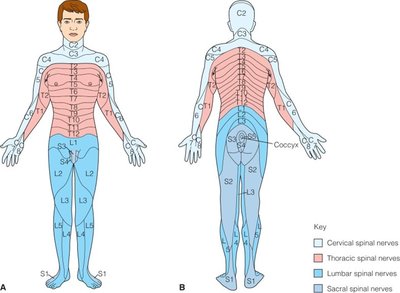
Reflexes
Somatic Reflexes: Involve skeletal muscle contraction, mediated by the spinal cord.
Autonomic Reflexes: Activate cardiac muscle, smooth muscle, and glands.
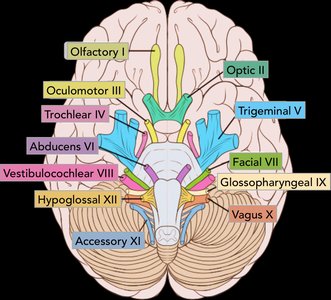
Cranial Nerves
There are 12 cranial nerves, each with specific functions related to head, neck, and some body regions.
Mnemonic: "Oh, Oh, Oh, To Touch And Feel Very Good Velvet, Ah!"
Assessment: Testing cranial nerve function is essential in neurological evaluation.
Autonomic Nervous System
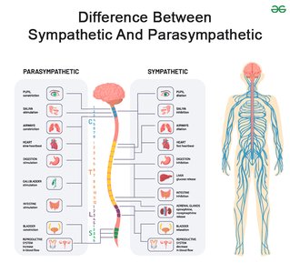
The autonomic nervous system regulates homeostasis and is divided into:
Sympathetic Nervous System: Stress response, "fight or flight".
Parasympathetic Nervous System: Resting state, "rest and digest".
Cerebral Circulation and Blood-Brain Barrier
Cerebral Artery Circulation
The brain is supplied by the internal carotid and vertebral arteries, which unite in the Circle of Willis to provide collateral circulation. 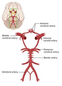
Blood-Brain Barrier
The blood-brain barrier is a selective permeability system that protects the brain from toxins and pathogens while allowing essential nutrients to pass. 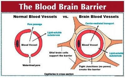
Intracranial Pressure (ICP) and Cerebral Perfusion Pressure (CPP)
Monro-Kellie Hypothesis
The Monro-Kellie Hypothesis states that the total volume of brain tissue, CSF, and blood within the cranium must remain constant. Changes in one component must be compensated by changes in another to maintain normal ICP. 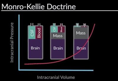
Cerebral Perfusion Pressure (CPP)
CPP is the pressure gradient required to supply blood to the brain, calculated as the difference between mean arterial pressure (MAP) and ICP.
Formula:
Normal CPP: 70–80 mmHg
Ischemia: Occurs at CPP of 30–40 mmHg
Irreversible Damage: CPP < 25 mmHg

Neurological Assessment
Glasgow Coma Scale (GCS)
The GCS is a standardized tool for assessing level of consciousness, with scores ranging from 3 (deep coma) to 15 (fully alert).
Components: Eye opening, verbal response, motor response.
Score < 8: Indicates need for airway protection.
Posturing
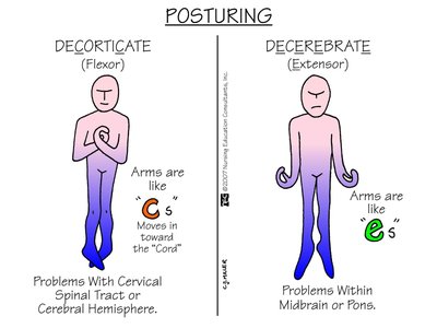
Decorticate: Abnormal flexion, indicates cerebral hemisphere dysfunction.
Decerebrate: Abnormal extension, indicates brainstem dysfunction.
Pupil Assessment
Pupillary reactions help assess neurological function and possible lesions.
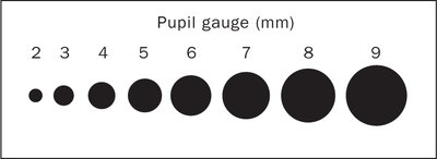
Size: Standardized pupil gauge used for comparison.
Symmetry: Unequal pupils may indicate neurological change.
Reaction to Light: Non-reactive pupils may indicate trauma or drug effects.
Common Neurological Disorders
Cerebral Vascular Accident (CVA) / Stroke
Ischemic Stroke: Caused by thrombus or embolism, treated with thrombolytics if no hemorrhage.
Hemorrhagic Stroke: Caused by ruptured aneurysm or trauma, requires neurosurgical intervention.
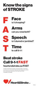
Assessment: Early recognition and treatment are critical.
Transient Ischemic Attack (TIA)
TIA is a temporary interruption of cerebral blood flow, resulting in brief neurological deficits without permanent injury.
Wernicke Encephalopathy
Acute neurological disorder caused by thiamine deficiency, often related to alcoholism and malnutrition.
Delirium vs Dementia
Delirium: Acute, fluctuating mental status, attention deficits, and altered LOC.
Dementia: Chronic, progressive cognitive decline.
Seizures
Seizures are caused by abnormal electrical activity in the brain and are classified as generalized or partial.
Tonic-Clonic: Two phases: tonic (rigid muscles) and clonic (alternating contraction/relaxation).
Status Epilepticus: Prolonged seizure > 5 mins, requires rapid intervention.
Guillan-Barre Syndrome
Autoimmune disorder causing rapid-onset muscle weakness, often triggered by infection or vaccination.
Degenerative Neurological Disorders
Parkinson's Disease: Dopamine deficiency, tremor, rigidity, postural instability.
Multiple Sclerosis: Progressive demyelination, exacerbations and remissions.
Myasthenia Gravis: Weakness due to impaired nerve-muscle communication.
Huntington's Disease: Genetic disorder, chorea, dementia.
Traumatic Brain and Spinal Cord Injuries
Traumatic Brain Injury (TBI)
TBI results from mechanical disruption of brain tissue, ranging from mild (concussion) to severe (hematoma, diffuse axonal injury).
Spinal Cord Injury (SCI)
SCI symptoms depend on injury location and extent. Cervical injuries can cause quadriplegia; lower injuries cause paraplegia.
Complications: Respiratory issues, autonomic dysreflexia, abnormal perfusion, bladder/bowel dysfunction, neuropathic pain.
Example: Autonomic dysreflexia is a life-threatening complication in SCI at or above T6, triggered by noxious stimuli below the injury level.
Summary Table: CNS vs PNS Neuroglia
Central Nervous System | Peripheral Nervous System |
|---|---|
Ependymal cells, Oligodendrocytes, Astrocytes, Microglia | Satellite cells, Schwann cells |
Additional info: Academic context was added to clarify the functions and clinical relevance of each structure and disorder.
