 뒤로
뒤로Respiratory System: Pulmonary Function, Gas Exchange, and Clinical Assessment
스터디 가이드 - 스마트 노트
 자료에 맞춘 맞춤형 노트, 핵심 정의, 예시, 맥락을 확장해 제공합니다.
Respiratory System Overview
Anatomy of the Respiratory System
The respiratory system is essential for gas exchange, supplying oxygen to the body and removing carbon dioxide. It consists of upper and lower airways, lungs, and associated structures.
Upper Airways: Nose, mouth, pharynx, larynx
Lower Airways: Trachea, bronchi, bronchioles, alveoli
Muscles: Diaphragm, intercostal muscles, abdominal muscles
Thoracic Cage: Ribs
Pleura: Parietal and visceral layers
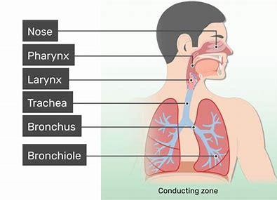
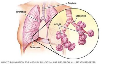
Conducting Airways
Conducting airways move air to and from the atmosphere and lungs. They humidify, filter, and warm air, and contain a mucociliary system to remove pathogens and foreign materials.
Mucociliary System: Captures pathogens in mucus and transports them to the pharynx for elimination.
Non-sterile Environment: Upper airways are non-sterile, important for nursing care.
Mechanics of Ventilation
Ventilation Process
Ventilation is the movement of air from the atmosphere to the alveoli, driven by contraction and relaxation of the diaphragm and intercostal muscles.
Inspiration: Thorax expands, intrapulmonary pressure decreases, air flows in.
Expiration: Thorax relaxes, pressure increases, air flows out.
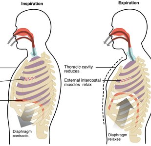
Lung Compliance
Lung compliance measures the ease of lung expansion.
High Compliance: Lungs stretch easily, optimal gas exchange.
Low Compliance: "Stiff lungs" require more pressure to inflate, seen in restrictive lung diseases.
PEEP (Positive End Expiratory Pressure): Used to keep alveoli open.
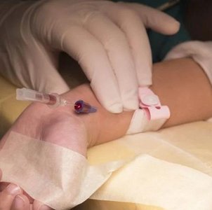
Lung Inflation and Surfactant
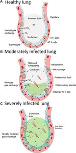
Surfactant: Lipoprotein secreted by type II alveolar cells reduces surface tension, preventing alveolar collapse.
Thoracic Structure: Rib cage and pleura help maintain lung inflation.
Gas Exchange and Diffusion
Pulmonary Gas Exchange
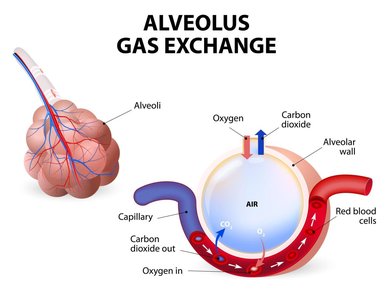
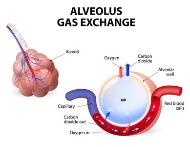
Gas exchange occurs at the alveolar-capillary membrane, where oxygen diffuses into blood and carbon dioxide diffuses out.
External Respiration: Oxygen moves from alveoli to pulmonary capillaries; CO2 moves from capillaries to alveoli.
Internal Respiration: Oxygen moves from blood to cells; CO2 moves from cells to blood.
Factors Affecting Diffusion
Partial Pressures: PaO2, PaCO2, PvO2, PvCO2
Pressure Gradient: Drives direction and rate of gas movement
Surface Area: Greater area increases gas exchange
Thickness of Membrane: Increased thickness decreases diffusion (e.g., pulmonary edema, fibrosis)
Time: Exposure time for diffusion, affected by cardiac output
Oxygen Transport and Oxyhemoglobin Dissociation Curve
Hemoglobin and Oxygen Transport
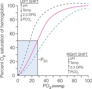
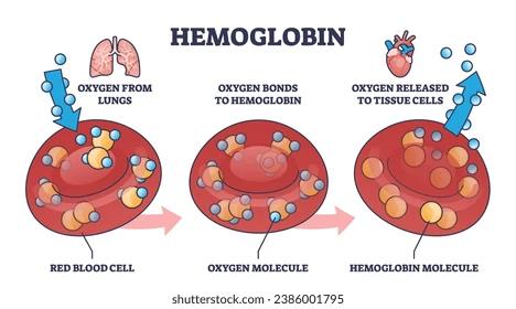
Hemoglobin (Hgb) binds oxygen in the lungs and releases it to tissues. The oxyhemoglobin dissociation curve illustrates the relationship between O2 bound to Hgb and partial pressure of O2.
Left Shift: Increased affinity, O2 tightly bound, less released to tissues (causes: alkalosis, hypothermia, hypocapnia, decreased 2,3-DPG)
Right Shift: Decreased affinity, O2 loosely bound, more released to tissues (causes: acidosis, hyperthermia, hypercapnia, increased 2,3-DPG)

Pulmonary Perfusion and V/Q Relationship
Pulmonary Perfusion
Blood is pumped from the right heart to the lungs for oxygenation, then returned to the left heart for systemic circulation.
Factors: Hemoglobin concentration, affinity of O2 to Hgb, gravity, cardiac output, blood flow, vasoconstriction
Ventilation-Perfusion (V/Q) Ratio
The V/Q ratio compares alveolar ventilation (V) to pulmonary capillary perfusion (Q).
Normal Ratio: 0.8 (4L air : 5L blood)
V/Q Mismatch: Seen in pulmonary embolism, shunting, and other disorders
Pulmonary Shunting
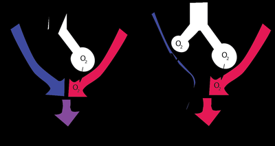
Pulmonary shunting refers to blood bypassing gas exchange, contributing to hypoxemia.
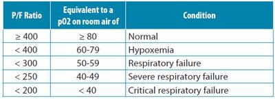
P/F Ratio | Equivalent pO2 (room air) | Condition |
|---|---|---|
≥ 400 | ≥ 80 | Normal |
< 400 | 60-79 | Hypoxemia |
< 300 | 50-59 | Respiratory failure |
< 250 | 40-49 | Severe respiratory failure |
< 200 | < 40 | Critical respiratory failure |
Ventilation Changes in the Older Adult
Age-Related Changes
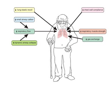
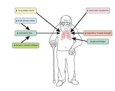
Aging affects the respiratory system, decreasing lung compliance and gas exchange efficiency.
Flattened diaphragm
Stiffened chest wall
Increased rib cage diameter
Weakened respiratory muscles
Air trapping and decreased gas exchange
Assessment and Diagnostics
Respiratory Assessment
Vital Signs: Pulse, BP, respiratory rate, temperature
Pulse Oximetry (SpO2): Measures oxygen saturation
Capnography: Measures exhaled CO2 (etCO2)
Physical Exam: Inspection, palpation, percussion, auscultation
Pulmonary Function Tests (PFTs)
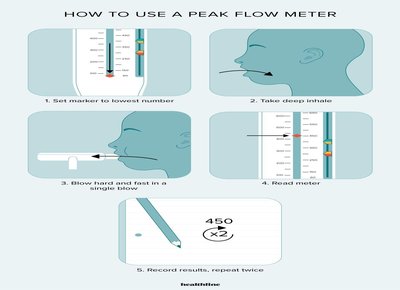
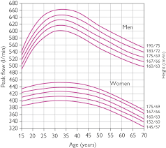
PFTs assess ventilation, muscle strength, and disease progression.
Tidal Volume: Air moved in/out per breath
Vital Capacity: Maximum air expired after maximal inspiration
Minute Ventilation: Total air expired in 1 minute
Common Pulmonary Disorders
Restrictive Pulmonary Disorders
Restrictive lung diseases limit lung expansion, reducing total lung capacity and oxygenation.
Intrinsic: Pulmonary fibrosis, sarcoidosis, occupational lung diseases
Extrinsic: Neuromuscular diseases, obesity
Obstructive Pulmonary Disorders
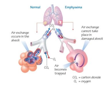
Obstructive diseases restrict airflow, causing air trapping and hypercapnia.
Examples: COPD, bronchitis, asthma
Symptoms: Dyspnea, wheezing, cough
Respiratory Medications
Beta Agonists
Short-acting: Salbutamol (Ventolin), Albuterol
Action: Bronchodilation via beta-2 adrenergic stimulation
Adverse Effects: Tachycardia, hypokalemia, hypertension, tremors
Anticholinergic Bronchodilators
Examples: Ipratropium bromide (Atrovent), Tiotropium (Spiriva)
Action: Blocks cholinergic-induced bronchoconstriction
Side Effects: Cough, dry mouth
Corticosteroids
Forms: IV, PO, inhaled
Action: Anti-inflammatory, reduces airway inflammation
Side Effects: Delayed wound healing, GI upset, immune suppression, oral thrush (inhaled)
Acute Respiratory Failure and ARDS
Acute Respiratory Failure
Occurs when the cardiopulmonary system cannot meet oxygen demands, resulting in hypoxia and/or hypercapnia.
Type 1: Hypoxemic (PaO2 ↓)
Type 2: Hypercarbic (PaCO2 ↑)
Symptoms: Dyspnea, tachypnea, cyanosis, altered LOC
Acute Respiratory Distress Syndrome (ARDS)
ARDS is a diffuse lung injury with severe inflammation and fluid accumulation, impairing gas exchange.
Triggers: Pneumonia, sepsis, COVID-19
Diagnostic Criteria: Rapid onset, bilateral opacities, PaO2/FiO2 < 300 mmHg
Interventions: Neuromuscular blocking agents, prone positioning, ECMO
Pulmonary Embolism and Respiratory Infections
Pulmonary Embolism (PE)
PE is a blood clot blocking pulmonary vasculature, restricting blood flow and gas exchange.
Risk Factors: Immobilization, surgery, smoking, malignancy, pregnancy
Diagnosis: CT angiogram, ultrasound
Interventions: Anticoagulants, thrombolysis, IVC filter
Respiratory Infections
Aspiration Pneumonia: Oral/gastric secretions reach lungs
Viral Pneumonia: Influenza, RSV, coronavirus
Bacterial Pneumonia: Streptococcus pneumoniae, MRSA, E. coli
Fungal Pneumonia: Aspergillus, Candida (immunocompromised)
Chest Trauma and Pleural Disorders
Rib Fractures and Flail Chest
Multiple rib fractures can cause flail chest, impairing ventilation and increasing risk for pneumonia and DVTs.
Pneumothorax
Pneumothorax is lung collapse due to loss of negative intrapleural pressure.
Types: Spontaneous, traumatic, tension
Interventions: Chest tube, needle decompression
Pleural Effusion, Hemothorax, Empyema
Pleural Effusion: Fluid accumulation in pleural space (transudative or exudative)
Hemothorax: Blood in pleural space
Empyema: Pus in pleural space
Interventions: Medications, chest tube
Summary Table: Key Pulmonary Terms
Term | Definition |
|---|---|
Ventilation | Movement of air in and out of lungs |
Diffusion | Movement of gases across alveolar-capillary membrane |
Perfusion | Blood flow through pulmonary capillaries |
Compliance | Ease of lung expansion |
PEEP | Positive End Expiratory Pressure |
ARDS | Acute Respiratory Distress Syndrome |
V/Q Ratio | Ventilation-perfusion ratio |
Pulse Oximetry | Measurement of oxygen saturation |
Capnography | Measurement of exhaled CO2 |
Formulas and Equations
Mean Arterial Pressure (MAP)
MAP is used to assess organ perfusion. The formula is:
P/F Ratio
P/F ratio is used to assess severity of respiratory failure:
Clinical Assessment and Nursing Interventions
Assess airway, breathing, circulation, disability (ABCD)
Monitor vital signs, SpO2, capnography
Support ventilation and oxygenation (e.g., supplemental O2, PEEP, BiPAP, CPAP)
Manage underlying conditions (infection, trauma, pulmonary disorders)
Positioning: "Good lung down" to improve V/Q matching
References
Wagner, K., Hardin-Pierce, M., & Welsh, D. (2018). High Acuity Nursing (7th ed.). Boston: Pearson.
Venus, K., Munshi, L. and Fralick, M. Prone positioning for the patient with hypoxic respiratory failure related to COVID-19: Canadian Medical Association Journal, 192(47), E1532-E1537
