 뒤로
뒤로Skeletal System: Structure, Function, and Medical Terminology
스터디 가이드 - 스마트 노트
 자료에 맞춘 맞춤형 노트, 핵심 정의, 예시, 맥락을 확장해 제공합니다.
Skeletal System Overview
Main Functions of the Skeletal System
The skeletal system is a complex network of bones and connective tissues that provides structural support, protection, and facilitates movement. It also serves as a reservoir for minerals and is essential for blood cell production.
Support: Framework for the body, maintaining shape and posture.
Protection: Shields vital organs (e.g., skull protects brain, rib cage protects heart and lungs).
Movement: Muscles attach to bones, enabling movement at joints.
Storage: Stores calcium ions (Ca2+), phosphate (PO43-), and lipids in bone matrix and marrow.
Blood Cell Production (Hematopoiesis): Occurs in red bone marrow.
Axial vs. Appendicular Skeleton
Classification of Bones
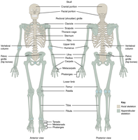
The skeleton is divided into two main regions: axial and appendicular. Understanding this classification is fundamental for medical terminology and anatomy.
Axial Skeleton: Includes bones of the head, neck, and trunk (skull, vertebral column, rib cage).
Appendicular Skeleton: Comprises bones of the limbs and girdles (shoulder, pelvic, arms, legs).
Bone Types: Compact vs. Spongy Bone
Characteristics and Locations
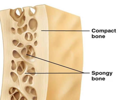
Bones are composed of two main types of tissue: compact and spongy bone, each with distinct structural and functional properties.
Compact Bone: Dense, forms the external layer of bones, organized in osteons, provides strength and rigidity.
Spongy Bone: Porous, found internally, consists of trabeculae, filled with red or yellow bone marrow, lighter and supports metabolic functions.
Structure of a Long Bone
Anatomical Features
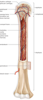
Long bones, such as the femur and humerus, have specialized structures that support their function and growth.
Epiphysis: Knob-like ends, site of articulation.
Diaphysis: Shaft, provides leverage and weight support.
Epiphyseal Plate/Line: Growth plate in youth (cartilage), becomes bone in adults.
Medullary Cavity: Central hollow area containing yellow marrow.
Articular Cartilage: Reduces friction at joints.
Periosteum: Fibrous membrane covering bone, contains blood vessels, nerves, and stem cells.
Red Bone Marrow Locations
Sites of Hematopoiesis
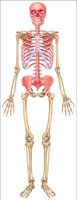
Red bone marrow is the primary site for blood cell production in adults, located in specific bones.
Found in spongy bone of axial skeleton, os coxae, and heads of femur and humerus.
Microscopic Structure of Bone
Osteons and Cellular Organization
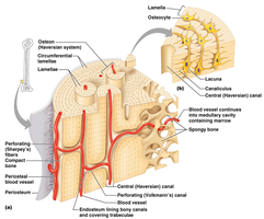
Bone tissue is organized into structural units called osteons, which facilitate nutrient delivery and waste removal.
Osteons: Cylindrical units running parallel to diaphysis.
Concentric Lamellae: Layers surrounding central canal.
Central Canal: Contains blood vessels and nerves.
Osteocytes: Mature bone cells in lacunae.
Canaliculi: Tiny channels for nutrient diffusion.
Microscopic Composition of Bone Tissue
Cell Types and Matrix Components
Bone tissue consists of specialized cells and a robust extracellular matrix, contributing to its mechanical properties.
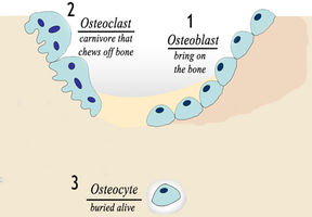
Osteoblasts: Build bone matrix (deposition).
Osteocytes: Maintain and repair bone.
Osteoclasts: Break down bone matrix (resorption), derived from white blood cells.
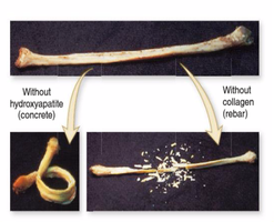
Ground Substance: Calcium phosphate salts (inorganic, resist compression).
Protein Fibers: Collagen (organic, provides flexibility and tensile strength).
Bone Remodeling
Bone remodeling is a continuous process involving osteoblasts and osteoclasts, replacing old bone tissue every 4-10 years.
Diet, Exercise, and Bone Remodeling
Wolff’s Law and Bone Health
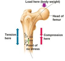
Bone adapts to mechanical stress according to Wolff’s Law, and its health is influenced by diet and activity.
Wolff’s Law: Osteoblasts reinforce stressed areas, making bones stronger with regular exercise.
Calcium: Essential for bone matrix.
Vitamin D: Facilitates calcium absorption.
Protein: Required for collagen synthesis.
Osteoporosis
Pathology and Risk Factors
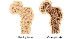
Osteoporosis is a disorder characterized by decreased bone mass and increased fracture risk, especially in postmenopausal women.
Causes: Hormonal changes, inactivity, poor diet.
Effects: Weakened bone structure, higher likelihood of fractures.
Bone Fracture Repair
Stages of Healing
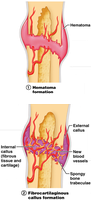
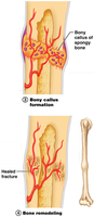
Bone fracture repair is a multi-stage process involving inflammation, callus formation, and remodeling.
Inflammation and Hematoma Formation: Blood vessels rupture, forming a hematoma; bone tissue dies.
Cartilaginous Callus Formation: Stem cells produce cartilage, bridging the fracture.
Bony Callus Formation: Cartilage is replaced by bone matrix.
Bone Remodeling: Osteoclasts and osteoblasts reshape bone to its original form.
Joints (Articulations)
Types of Joints
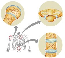
Joints are sites where two bones meet, classified by their structure and movement capacity.
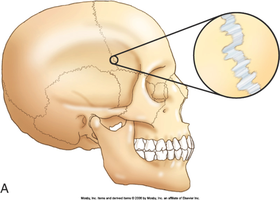
Fibrous Joints: Held by fibrous tissue, allow no movement (e.g., sutures in skull).
Cartilaginous Joints: Held by cartilage, allow slight movement (e.g., intervertebral discs).
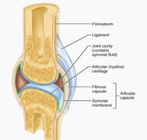
Synovial Joints: Fluid-filled cavity, most common, allow full movement, stabilized by ligaments and tendons.
Structure of Synovial Joints
Synovial joints have specialized structures to facilitate movement and reduce friction.
Fibrous Capsule: Encloses the joint.
Articular Cartilage: Smooth surface covering bone ends.
Synovial Membrane: Secretes synovial fluid.
Joint Cavity: Contains synovial fluid.
Synovial Joint Movements
Types of Movements
Synovial joints allow a variety of movements, each with specific terminology.
Flexion: Decreases angle between bones.
Extension: Increases angle between bones.
Adduction: Moves joint toward midline.
Abduction: Moves joint away from midline.
Elevation/Depression: Moves joint superiorly/inferiorly (e.g., mandible, shoulder).
Sprains, Dislocations, and Arthritis
Joint Disorders
Common joint disorders include sprains, dislocations, and arthritis, each affecting joint stability and function.
Sprain: Ligament is stretched or torn.
Dislocation: Bones are moved out of normal position due to ligament injury.
Osteoarthritis: Wear and tear of articular cartilage.
Rheumatoid Arthritis: Autoimmune disease affecting synovial membrane.
Summary Table: Bone Cell Types and Functions
Cell Type | Function |
|---|---|
Osteoblast | Builds bone matrix (deposition) |
Osteocyte | Maintains and repairs bone |
Osteoclast | Breaks down bone matrix (resorption) |
Summary Table: Joint Types
Joint Type | Structure | Movement | Example |
|---|---|---|---|
Fibrous | Fibrous connective tissue | None | Sutures in skull |
Cartilaginous | Cartilage | Slight | Intervertebral discs |
Synovial | Fluid-filled cavity | Full | Knee, shoulder |
Key Medical Terminology
Hematopoiesis: Blood cell formation in red bone marrow.
Epiphysis: End of a long bone.
Diaphysis: Shaft of a long bone.
Osteon: Structural unit of compact bone.
Trabeculae: Rod-like structures in spongy bone.
Articulation: Joint where two bones meet.
Relevant Equations
Bone matrix composition:
Inorganic component: (calcium phosphate)
Organic component: Collagen fibers
Bone remodeling rate:
Complete replacement every 4-10 years
Additional info: Tables and explanations were expanded for academic completeness and clarity. Images included only when directly relevant to the adjacent content.
