 뒤로
Chapter 5: Eukaryotic Cells and Microorganisms: Structure, Function, and Diversity- Book Notes
스터디 가이드 - 스마트 노트
 자료에 맞춘 맞춤형 노트, 핵심 정의, 예시, 맥락을 확장해 제공합니다.
Overview of Eukaryotes
Evolutionary Origins and Endosymbiosis
Eukaryotic cells are believed to have evolved from a common ancestor shared with bacteria and archaea. The process of endosymbiosis played a critical role in the development of complex eukaryotic cells, particularly through the incorporation of mitochondria and chloroplasts, which originated from free-living bacteria.
Last Common Ancestor (LCA): The precursor cell from which bacteria, archaea, and eukaryotes diverged.
Endosymbiosis: The theory that mitochondria and chloroplasts were once independent prokaryotic organisms that became permanent residents within ancestral eukaryotic cells.
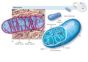
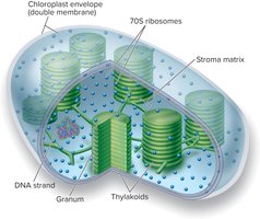
Evidence for Endosymbiosis: Mitochondria and chloroplasts contain their own circular DNA, 70S ribosomes, and double membranes.
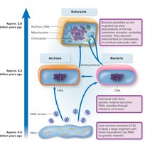
Types of Eukaryotic Microorganisms: Includes protozoa (unicellular), fungi (unicellular and multicellular), and algae (unicellular and multicellular). Animals and plants are also eukaryotic but are not typically studied as microorganisms.
Form and Function of the Eukaryotic Cell: External and Boundary Structures
General Features of Eukaryotic Cells
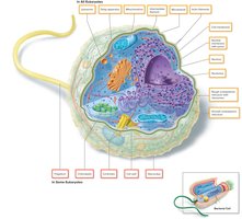
Eukaryotic cells are characterized by the presence of membrane-bound organelles and a complex internal structure. All eukaryotic cells possess a cytoplasmic membrane, nucleus, mitochondria, endoplasmic reticulum, Golgi apparatus, vacuoles, cytoskeleton, and glycocalyx. Some groups also have cell walls, locomotor appendages, and chloroplasts.
Flagella and Cilia
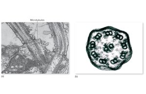
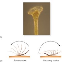
Eukaryotic Flagella: Thicker and more complex than prokaryotic flagella, with a 9+2 arrangement of microtubules. They are covered by the cell membrane and move in a whipping motion.
Cilia: Shorter and more numerous than flagella, found in certain protozoa and animal cells. They function in locomotion and feeding, moving with a coordinated oarlike motion.
The Glycocalyx
The glycocalyx is an outermost boundary layer composed of polysaccharides, functioning as a protective extracellular matrix. It can appear as a network of fibers, a slime layer, or a capsule.
Cell Wall
Present in fungi and algae, the eukaryotic cell wall provides structural support and shape. Unlike bacterial cell walls, fungal cell walls are composed of chitin or cellulose and have a different chemical structure.
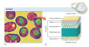
Cytoplasmic Membrane
Composed of a phospholipid bilayer with embedded proteins and sterols for membrane stability.
Acts as a selectively permeable barrier with advanced transport mechanisms for nutrients and waste.
Form and Function of the Eukaryotic Cell: Internal Structures
The Nucleus
The nucleus is the control center of the cell, containing the genetic material (DNA) organized as chromatin. It is surrounded by a double-layered nuclear envelope with pores for molecular exchange. The nucleolus within the nucleus is the site of ribosomal RNA synthesis.
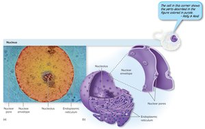
Mitosis and Meiosis
Mitosis: The process of cell division resulting in two genetically identical daughter cells.
Meiosis: Specialized cell division producing sex cells (gametes) with half the chromosome number.
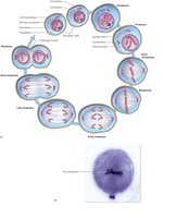
Endoplasmic Reticulum (ER)
Rough ER (RER): Studded with ribosomes; involved in protein synthesis and transport.
Smooth ER (SER): Lacks ribosomes; involved in synthesis and storage of lipids and other nonprotein molecules.
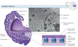
Golgi Apparatus
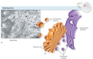
The Golgi apparatus modifies, sorts, and packages proteins and lipids for secretion or delivery to other organelles. It consists of stacked, flattened sacs called cisternae and works closely with the ER via vesicular transport.
Vesicular Transport
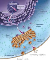
Proteins synthesized in the RER are transported to the Golgi apparatus in transitional vesicles, modified, and then sent to their destinations in condensing vesicles. This process is essential for secretion and intracellular trafficking.
Lysosomes and Vacuoles
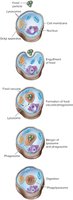
Lysosomes: Contain digestive enzymes for intracellular digestion and defense against pathogens.
Vacuoles: Membrane-bound sacs for storage, digestion, and excretion of substances.
Mitochondria
Mitochondria are the energy generators of the cell, producing ATP through aerobic respiration. They have an outer membrane, a highly folded inner membrane (cristae), and a matrix containing enzymes, DNA, and 70S ribosomes. Mitochondria divide independently and are evidence of endosymbiotic origin.
Chloroplasts
Chloroplasts are found in algae and plants, enabling photosynthesis. They contain thylakoids (stacked into grana), stroma matrix, DNA, and 70S ribosomes, supporting their endosymbiotic origin.
Ribosomes
Sites of protein synthesis, found free in the cytoplasm, attached to the RER, or within mitochondria and chloroplasts.
Eukaryotic ribosomes are 80S (composed of 60S and 40S subunits), while prokaryotic ribosomes are 70S.
The Cytoskeleton
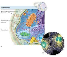
The cytoskeleton provides structural support, facilitates intracellular transport, and enables cell movement. It consists of three main types of fibers: actin filaments, intermediate filaments, and microtubules.
The Fungi
General Features and Morphology
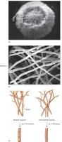
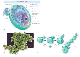
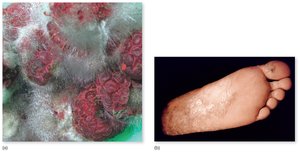
Fungi are a diverse group of eukaryotic organisms, including macroscopic forms (mushrooms) and microscopic forms (molds and yeasts). Yeasts are unicellular, round to oval, and reproduce asexually by budding. Molds consist of long, threadlike hyphae that form a mycelium. Some fungi are dimorphic, switching between yeast and mold forms depending on environmental conditions.
Fungal Nutrition
Heterotrophic: Obtain nutrients from organic sources.
Saprobes: Decompose dead organic matter.
Parasites: Derive nutrients from living hosts.
Organization and Reproduction
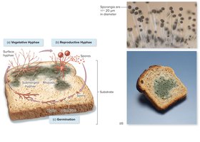
Mycelium: Intertwined mass of hyphae forming the body of a mold.
Septa: Cross-walls dividing hyphae into segments.
Spores: Reproductive bodies, produced asexually or sexually.
Asexual Spore Formation
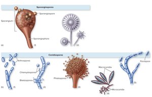
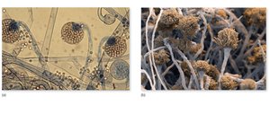
Sporangiospores: Formed within a sac (sporangium).
Conidiospores (Conidia): Free spores not enclosed in a sac.
Fungal Identification and Medical Importance
Identification is based on spore type, hyphal structure, colony characteristics, and genetic analysis.
Fungi can cause infections (mycoses), allergies, and produce toxins. They also have beneficial roles in decomposition, symbiosis with plants, and production of antibiotics and food products.
The Protists
Algae
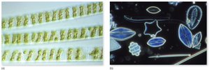
Algae are photosynthetic protists found in aquatic environments. They possess chloroplasts and contribute significantly to global oxygen production. Some algae can produce toxins harmful to humans and animals.
Protozoa
Single-celled eukaryotes lacking true tissues.
Exhibit diverse forms and specialized organelles for feeding, movement, and reproduction.
Most are free-living; some are important human parasites.
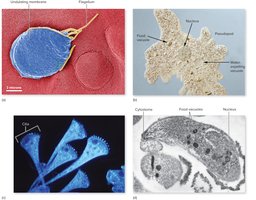
Protozoan Locomotion
Pseudopods: Amoeboid movement and feeding structures.
Flagella: One or more long, whip-like structures.
Cilia: Numerous short, hair-like structures covering the cell.
Life Cycle and Reproduction
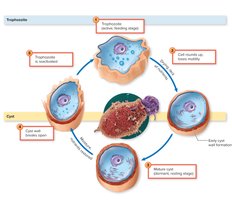
Trophozoite: Active, feeding stage.
Cyst: Dormant, resistant stage formed under adverse conditions; important for transmission of disease.
Reproduction is mainly asexual (mitosis), but sexual processes (conjugation) also occur in some groups.
The Helminths
Major Groups and Morphology
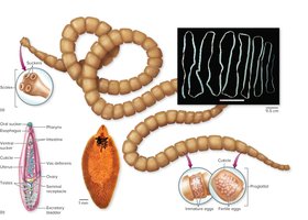
Flatworms: Thin, segmented (cestodes/tapeworms) or unsegmented (trematodes/flukes).
Roundworms (Nematodes): Cylindrical, unsegmented worms.
Helminths are multicellular with developed organ systems, especially reproductive systems.
Life Cycles and Transmission
Typical stages: fertilized egg, larva, adult.
Transmission can occur via food, water, soil, or vectors. Humans may serve as definitive or intermediate hosts.
Some helminths are hermaphroditic, others have separate sexes.
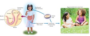
Example: Pinworm Life Cycle
Enterobius vermicularis is a common intestinal roundworm. Infection occurs via ingestion of eggs, which hatch and mature in the intestine. Eggs are released and can be transmitted to new hosts through contaminated hands or surfaces.
Identification and Importance
Helminths are identified by morphology, reproductive structures, and egg/larval characteristics.
They are globally distributed and can cause significant morbidity, especially in tropical regions.
Absence of helminth infections in developed countries may be linked to increased autoimmune and allergic diseases.
