 뒤로
Functional Anatomy of Prokaryotic and Eukaryotic Cells: Microbiology Study Notes
스터디 가이드 - 스마트 노트
 자료에 맞춘 맞춤형 노트, 핵심 정의, 예시, 맥락을 확장해 제공합니다.
Functional Anatomy of Prokaryotic and Eukaryotic Cells
Comparing Prokaryotic and Eukaryotic Cells
Understanding the structural and functional differences between prokaryotic and eukaryotic cells is fundamental in microbiology. These differences influence cellular processes, pathogenicity, and classification.
Prokaryotes: Cells lacking a membrane-bound nucleus. Usually possess a single circular chromosome, no histones, and lack membrane-enclosed organelles. Bacteria have peptidoglycan cell walls; archaea have pseudomurein cell walls. Divide by binary fission.
Eukaryotes: Cells with a true nucleus enclosed by a nuclear membrane. Possess paired chromosomes, histones, and membrane-bound organelles. Cell walls, when present, are made of polysaccharides. Divide by mitosis.
The Size, Shape, and Arrangement of Bacterial Cells
Bacteria exhibit a variety of shapes and arrangements, which are important for identification and classification.
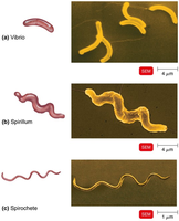
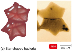
Shapes: Bacillus (rod-shaped), coccus (spherical), spiral (vibrio, spirillum, spirochete), star-shaped, and rectangular.
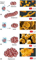
Arrangements: Pairs (diplococci, diplobacilli), clusters (staphylococci), chains (streptococci, streptobacilli), tetrads (groups of four), sarcinae (cubelike groups of eight).
Monomorphic: Most bacteria have a single shape.
Pleomorphic: Some bacteria can vary in shape.
Example: Bacillus is both a scientific name and a shape descriptor.
Structures External to the Cell Wall
External structures play key roles in protection, motility, and adherence.
Glycocalyx
External to the cell wall; viscous and gelatinous.
Made of polysaccharide and/or polypeptide.
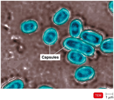
Capsule: Neatly organized, firmly attached; prevents phagocytosis and aids in adherence.
Slime layer: Unorganized, loose; helps form biofilms.
Contributes to virulence in pathogens such as Bacillus anthracis, Streptococcus pneumoniae, Klebsiella pneumoniae.
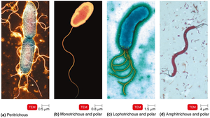
Flagella
Filamentous appendages for motility; made of flagellin protein.
Three parts: filament (outermost), hook (attaches filament), basal body (anchors flagellum).
Enable movement toward/away from stimuli (taxis); rotate for "run" or "tumble".
Flagella proteins are H antigens, used for serovar identification (e.g., Escherichia coli O157:H7).
Axial Filaments
Also called endoflagella; found in spirochetes.
Anchored at one end; rotation causes corkscrew movement.
Archaella
Motility structures in archaea; made of archaellins (glycoproteins).
Rotate like flagella; use ATP for energy.
Fimbriae and Pili
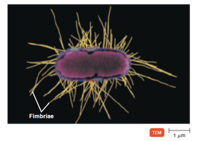
Fimbriae: Hairlike appendages for attachment; important in biofilm formation and adherence to surfaces (e.g., Neisseria gonorrhoeae, E. coli O157).
Pili: Involved in motility (gliding, twitching) and DNA transfer (conjugation).
The Cell Wall
The cell wall is essential for maintaining cell shape, preventing osmotic lysis, and contributing to pathogenicity. Its composition is a key factor in bacterial classification.
Made of peptidoglycan in bacteria; provides structural integrity.
Site of action for antibiotics (e.g., penicillin).
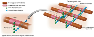
Peptidoglycan: Polymer of N-acetylglucosamine (NAG) and N-acetylmuramic acid (NAM), linked by polypeptides.
Penicillin disrupts peptide cross-bridges, weakening the cell wall.
Gram-Positive Cell Walls
Thick peptidoglycan layers.
Teichoic acids (lipoteichoic and wall teichoic acids) link cell wall to plasma membrane and peptidoglycan.
Carry negative charge; regulate cation movement.
High susceptibility to penicillin; disrupted by lysozyme.
Two rings in basal body of flagella; produce exotoxins.
Gram-Negative Cell Walls
Thin peptidoglycan layer; periplasmic space between outer and plasma membranes.
Outer membrane contains lipopolysaccharide (LPS), lipoproteins, and phospholipids.
LPS: O polysaccharide (antigenic), lipid A (endotoxin).
Porins form channels; protect from phagocytes, complement, antibiotics.
Low susceptibility to penicillin; four rings in basal body of flagella; produce endotoxins and exotoxins.
Atypical Cell Walls
Acid-fast cell walls: Similar to gram-positive, but with waxy mycolic acid. Stain with carbolfuchsin; genera include Mycobacterium and Nocardia.
Mycoplasmas: Lack cell walls; sterols in plasma membrane provide protection.
Archaea: Wall-less or walls of pseudomurein (lack NAM and D-amino acids).
Damage to the Cell Wall
Lysozyme hydrolyzes glycan bonds in peptidoglycan; penicillin inhibits peptide bridge formation.
Protoplast: Wall-less gram-positive cell.
Spheroplast: Wall-less gram-negative cell.
L forms: Wall-less cells with irregular shapes; susceptible to osmotic lysis.
Structures Internal to the Cell Wall
The Plasma (Cytoplasmic) Membrane
Phospholipid bilayer enclosing cytoplasm.
Contains peripheral, integral, and transmembrane proteins; some form channels.
Fluid mosaic model: proteins and phospholipids move freely; membrane is self-sealing.
Selective permeability; contains enzymes for ATP production; some have photosynthetic pigments (chromatophores).
Movement of Materials Across Membranes
Passive processes: No energy required; substances move from high to low concentration.
Simple diffusion: Solute moves until equilibrium is reached.
Facilitated diffusion: Transporter proteins enable movement of ions/large molecules.
Osmosis: Water moves across membrane from high to low concentration; through lipid layer or aquaporins.
Active transport: Requires transporter protein and ATP; moves substances against gradient.
Cytoplasm
Thick, aqueous, elastic substance inside plasma membrane.
Contains DNA (nucleoid), ribosomes, inclusions, cytoskeleton.
Cytoskeleton: fibers involved in cell division, shape, growth, DNA movement.
The Nucleoid
Bacterial chromosome: circular, double-stranded DNA; not membrane-bound.
Plasmids: small, extrachromosomal DNA circles; carry noncrucial genes (e.g., antibiotic resistance, toxin production); replicate independently; transferable.
Ribosomes
Sites of protein synthesis; made of protein and ribosomal RNA.
70S ribosomes in prokaryotes.
Antibiotics targeting ribosomes: streptomycin, gentamicin, erythromycin, chloramphenicol.
Inclusions
Reserve deposits of nutrients.
Types: metachromatic granules (phosphate), polysaccharide granules (energy), lipid inclusions (energy), sulfur granules (energy), carboxysomes (RuBisCO enzyme for photosynthesis), gas vacuoles (buoyancy), magnetosomes (iron oxide).
Endospores
Resting cells produced when nutrients are depleted; resistant to desiccation, heat, chemicals, radiation.
Survival mechanism; not reproductive.
Produced by Bacillus and Clostridium.
Sporulation: Endospore formation.
Germination: Endospore returns to vegetative state.
Important in food industry due to resistance.
Summary Table: Gram-Positive vs. Gram-Negative Bacteria
Feature | Gram-Positive | Gram-Negative |
|---|---|---|
Peptidoglycan | Thick | Thin |
Teichoic acids | Present | Absent |
Outer membrane | Absent | Present |
Flagella basal body | 2 rings | 4 rings |
Toxins | Exotoxins | Endotoxins & Exotoxins |
Penicillin susceptibility | High | Low |
Lysozyme susceptibility | High | Low |
Additional info: Table entries inferred from textbook context and standard microbiology knowledge.
