 뒤로
Functional Anatomy of Prokaryotic and Eukaryotic Cells
스터디 가이드 - 스마트 노트
 자료에 맞춘 맞춤형 노트, 핵심 정의, 예시, 맥락을 확장해 제공합니다.
Comparing Prokaryotic and Eukaryotic Cells
Overview of Cell Types
Cells are classified as either prokaryotic or eukaryotic based on structural and functional differences. The primary distinctions involve the presence or absence of a nucleus, organelles, and the composition of the cell wall and ribosomes.
Prokaryotes: Cells lacking a true nucleus and membrane-bound organelles. Includes Bacteria and Archaea.
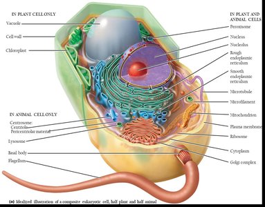
Eukaryotes: Cells with a true nucleus enclosed by a nuclear membrane and various membrane-bound organelles. Includes fungi, protozoa, algae, plants, and animals.
Feature | Prokaryotes | Eukaryotes |
|---|---|---|
Chromosomes | One circular, not in a membrane | Paired, in nuclear membrane |
Histones | Absent | Present |
Organelles | Absent | Present |
Cell Wall | Bacteria: peptidoglycan; Archaea: pseudomurein | Polysaccharide (when present) |
Division | Binary fission | Mitosis |
The Size, Shape, and Arrangement of Bacterial Cells
Bacterial Morphology
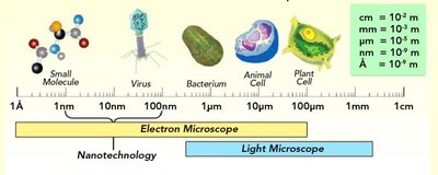
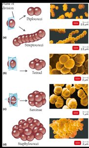
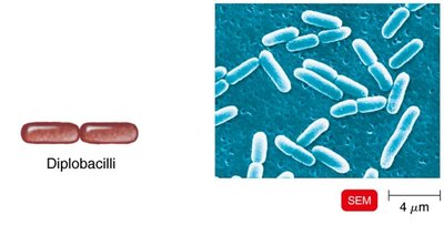
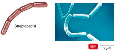
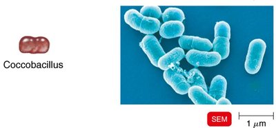
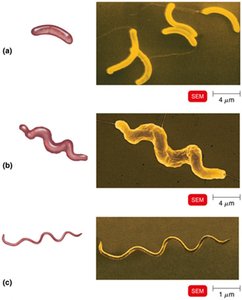
Bacteria exhibit a variety of shapes and arrangements, which are important for identification and classification. The average size of bacteria ranges from 0.2 to 2.0 μm in diameter and 2 to 8 μm in length.
Coccus: Spherical-shaped
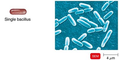
Bacillus: Rod-shaped
Spiral: Includes vibrio (comma-shaped), spirillum (rigid, corkscrew), and spirochete (flexible, helical)
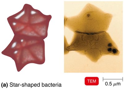
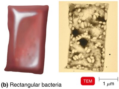
Other shapes: Star-shaped, rectangular
Structures External to the Cell Wall
Glycocalyx
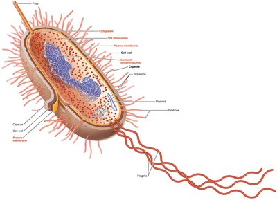
The glycocalyx is a viscous, gelatinous polymer external to the cell wall, composed of polysaccharide, polypeptide, or both. It can exist as a capsule (organized and firmly attached) or a slime layer (unorganized and loose). Capsules contribute to bacterial virulence and help in biofilm formation.
Capsule: Protects against phagocytosis and desiccation.
Slime layer: Facilitates attachment to surfaces.
Flagella
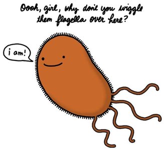
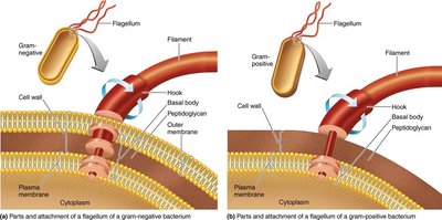
Flagella are long, filamentous appendages used for motility. They are composed of the protein flagellin and consist of three parts: filament, hook, and basal body. The arrangement and number of flagella can vary (peritrichous, monotrichous, lophotrichous, amphitrichous).
Function: Motility (taxis), antigenic variation (H antigen)
Axial Filaments
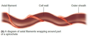
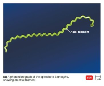
Axial filaments, also called endoflagella, are found in spirochetes. They are anchored at one end of the cell and cause movement in a corkscrew motion.
Fimbriae and Pili
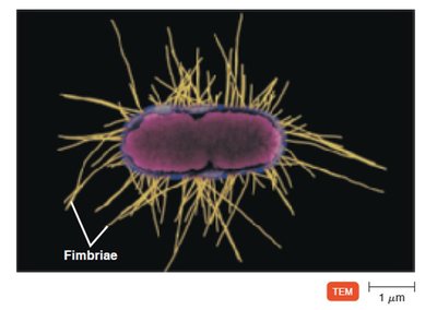
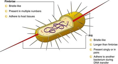
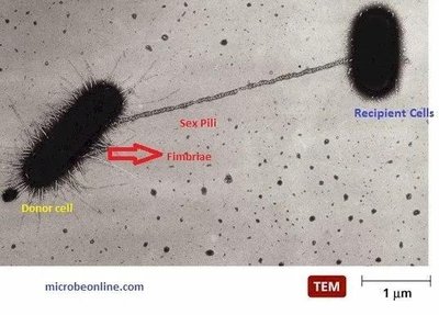
Fimbriae and pili are hairlike appendages found in many gram-negative bacteria. Fimbriae are numerous and aid in attachment and biofilm formation, while pili are longer, fewer, and involved in motility and DNA transfer (conjugation).
The Cell Wall
Structure and Function
The bacterial cell wall is a complex, semirigid structure that determines cell shape, protects against osmotic lysis, and serves as an anchorage point for flagella. It is also a target for certain antibiotics.
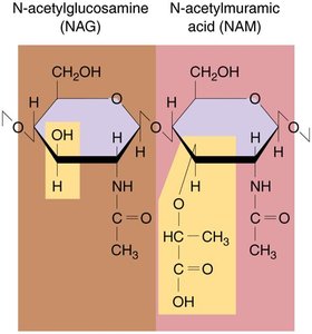
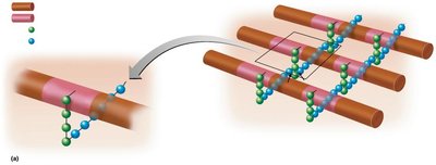
Main component: Peptidoglycan (murein), a polymer of N-acetylglucosamine (NAG) and N-acetylmuramic acid (NAM) linked by polypeptides.
Gram-Positive vs. Gram-Negative Cell Walls
Feature | Gram-Positive | Gram-Negative |
|---|---|---|
Peptidoglycan | Thick, multilayered | Thin, single layer |
Teichoic acids | Present | Absent |
Outer membrane | Absent | Present (contains LPS) |
Periplasmic space | Absent | Present |
Sensitivity to antibiotics | More sensitive | Less sensitive |
Atypical Cell Walls
Mycoplasma: Lack cell walls; plasma membrane contains sterols for protection.
Archaea: May lack cell walls or have walls of pseudomurein (no peptidoglycan).
Acid-fast bacteria: Mycobacterium and Nocardia have waxy mycolic acid in their cell walls, making them resistant to Gram staining.
Structures Internal to the Cell Wall
Plasma Membrane
The plasma membrane is a phospholipid bilayer with embedded proteins, responsible for selective permeability and containing enzymes for ATP production.
Cytoplasm
The cytoplasm is the substance inside the plasma membrane, consisting mainly of water, proteins, carbohydrates, lipids, ions, and a cytoskeleton.
Nucleoid
The nucleoid contains the bacterial chromosome (circular DNA) and may also contain plasmids, which carry non-essential genes such as antibiotic resistance.
Ribosomes
Prokaryotic ribosomes (70S) are the sites of protein synthesis and are composed of 50S and 30S subunits. They differ from eukaryotic ribosomes (80S) in size and density.
Endospores
Endospores are highly resistant, dormant structures formed by certain bacteria (e.g., Bacillus and Clostridium) in response to nutrient depletion. They are resistant to heat, desiccation, chemicals, and radiation.
Sporulation: Formation of endospores
Germination: Return to vegetative state
The Eukaryotic Cell
General Structure
Eukaryotic cells are larger and more complex than prokaryotic cells, containing a nucleus and various organelles. They include algae, protozoa, fungi, plants, and animals.
Flagella and Cilia
Flagella and cilia are projections used for locomotion or moving substances along the cell surface. Both are composed of microtubules arranged in a 9+2 pattern and move in a wavelike manner.
Cell Wall and Glycocalyx
The cell wall is present in plants, algae, and fungi, composed mainly of carbohydrates (cellulose, chitin, glucan, mannan). The glycocalyx is found in animal cells and is involved in cell-to-cell recognition and attachment.
Ribosomes
Eukaryotic ribosomes are 80S (60S + 40S subunits) and are found free in the cytoplasm or bound to the endoplasmic reticulum. 70S ribosomes are found in mitochondria and chloroplasts.
Nucleus
The nucleus is a double-membrane structure containing the cell's DNA complexed with histones to form chromatin, which condenses into chromosomes during cell division.
Mitochondria
Mitochondria are double-membraned organelles with inner folds (cristae) and matrix, responsible for ATP production through cellular respiration.
*Additional info: This guide covers the essential structural and functional differences between prokaryotic and eukaryotic cells, including specialized structures and their clinical significance. Tables and images are included to reinforce key concepts and aid in visual learning.*
