 뒤로
뒤로Functional Anatomy of Prokaryotic and Eukaryotic Cells: Microbiology Study Guide
스터디 가이드 - 스마트 노트
 자료에 맞춘 맞춤형 노트, 핵심 정의, 예시, 맥락을 확장해 제공합니다.
Functional Anatomy of Prokaryotic & Eukaryotic Cells
Principal Differences Between Prokaryotic and Eukaryotic Cells
Prokaryotic and eukaryotic cells are the two fundamental cell types in biology, each with distinct structural and functional characteristics. Understanding these differences is essential for microbiology students.
Chromosome: Prokaryotes typically have one circular chromosome, while eukaryotes possess paired chromosomes.
Nuclear Enclosure: Prokaryotes lack a nuclear membrane; eukaryotes have a well-defined nucleus.
Histones: Absent in prokaryotes, present in eukaryotes.
Membrane-Enclosed Organelles: Prokaryotes lack these; eukaryotes contain organelles such as mitochondria and endoplasmic reticulum.
Cell Division: Prokaryotes divide by binary fission; eukaryotes by mitosis.
Cell Walls: Prokaryotic cell walls are chemically complex (often peptidoglycan); eukaryotic cell walls, when present, are simpler (cellulose or chitin).
Flagella: Prokaryotic flagella are simple; eukaryotic flagella are complex, composed of microtubules.
Glycocalyx: Present as capsule or slime layer in prokaryotes; present in some eukaryotic cells lacking a cell wall.
Plasma Membrane: Prokaryotes generally lack sterols and carbohydrates; eukaryotes contain both.
Cytoplasm: Prokaryotes lack cytoskeleton and cytoplasmic streaming; eukaryotes possess both.
Ribosomes: Prokaryotes have 70S ribosomes; eukaryotes have 80S ribosomes in cytoplasm, 70S in mitochondria and chloroplasts.
Sexual Recombination: Prokaryotes transfer DNA only; eukaryotes involve meiosis.
Unique Characteristics of Prokaryotic Cells
Size, Shape, and Arrangement of Bacterial Cells
Bacteria exhibit a variety of sizes, shapes, and arrangements, which are important for identification and classification in microbiology.
Size: Average diameter is 0.2–2.0 µm, length 2–8 µm.
Shape: Most are monomorphic (single shape), but some are pleomorphic (variable shapes).
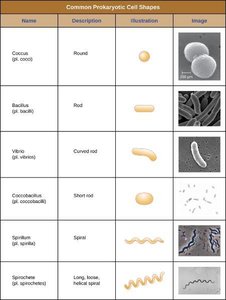
Common Shapes: Cocci (round), Bacilli (rod), Vibrio (curved rod), Coccobacillus (short rod), Spirillum (spiral), Spirochete (long, flexible spiral).
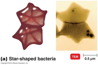
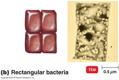
Unusual Shapes: Star-shaped and rectangular bacteria also exist.
Name | Description | Illustration | Image |
|---|---|---|---|
Coccus | Round | Illustration | Image |
Bacillus | Rod | Illustration | Image |
Vibrio | Curved rod | Illustration | Image |
Coccobacillus | Short rod | Illustration | Image |
Spirillum | Spiral | Illustration | Image |
Spirochete | Long, loose, helical spiral | Illustration | Image |
Arrangement of Bacterial Cells
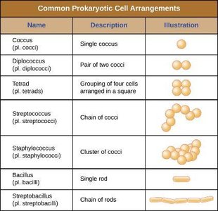
Bacterial cells cluster in patterns based on their plane of division, which aids in identification.
Name | Description | Illustration |
|---|---|---|
Coccus | Single coccus | Illustration |
Diplococcus | Pair of two cocci | Illustration |
Tetrad | Grouping of four cells arranged in a square | Illustration |
Streptococcus | Chain of cocci | Illustration |
Staphylococcus | Cluster of cocci | Illustration |
Bacillus | Single rod | Illustration |
Streptobacillus | Chain of rods | Illustration |
Prokaryotic Cell Structures & Functions
Plasma Membrane Structure
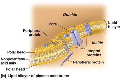
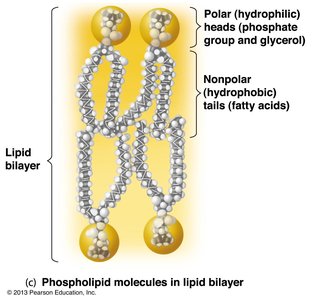
The plasma membrane is a selectively permeable barrier composed of a phospholipid bilayer with embedded proteins. It separates the intracellular and extracellular environments.
Phospholipid Bilayer: Consists of polar (hydrophilic) heads and nonpolar (hydrophobic) tails.
Proteins: Peripheral, integral, and transmembrane proteins serve various functions including transport and signaling.
Fluid Mosaic Model: Describes the dynamic arrangement of lipids and proteins.
Selective Permeability and Membrane Function
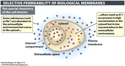
The plasma membrane maintains distinct chemical environments inside and outside the cell, allowing selective transport of substances.
Selective Permeability: Some substances (e.g., Na+) are abundant outside, others (e.g., K+) inside.
Transport Mechanisms: Include passive and active transport.
Passive Transport: Diffusion and Facilitated Diffusion
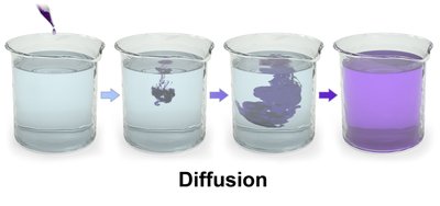
Passive transport involves the movement of substances down their concentration gradient without energy input.
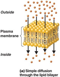
Simple Diffusion: Substances pass directly through the membrane.
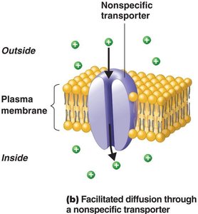
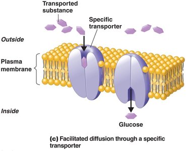
Facilitated Diffusion: Substances move via specific transporter proteins.

Osmosis and Osmotic Pressure
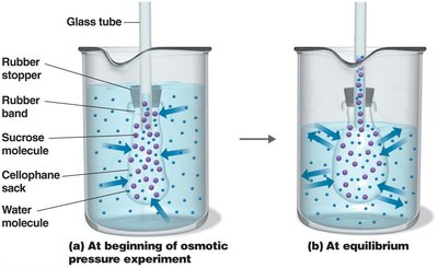
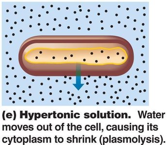
Osmosis is the movement of water across a semipermeable membrane from an area of low solute concentration to high solute concentration. Osmotic pressure is the force required to prevent water movement.
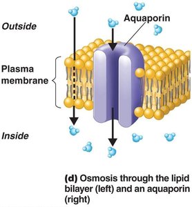
Osmosis: Water moves through the lipid bilayer or via aquaporins.
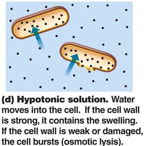
Osmotic Pressure: Determines cell stability in different solutions.
Active Transport
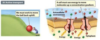
Active transport is the movement of substances against their concentration gradient, requiring energy input (usually ATP).
Mechanism: Utilizes membrane proteins and energy to move molecules.
Example: Sodium-potassium pump in cells.
Passive Transport
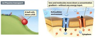
Passive transport is the spontaneous movement of substances down their concentration gradient without energy input.
Mechanism: Includes diffusion and facilitated diffusion.
Cytoplasm and Internal Structures
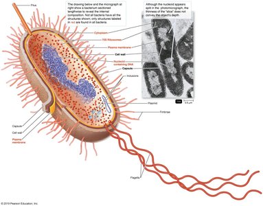
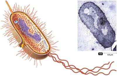
The cytoplasm is the substance inside the plasma membrane, consisting of water, proteins, carbohydrates, lipids, and ions. It contains various internal structures essential for cell function.
Nucleoid: Contains the bacterial chromosome, lacks nuclear envelope and histones.
Plasmids: Extrachromosomal genetic elements carrying non-crucial genes (e.g., antibiotic resistance).
Ribosomes: Sites of protein synthesis; prokaryotes have 70S ribosomes.
Inclusions: Reserve deposits such as metachromatic granules, polysaccharide granules, lipid inclusions, sulfur granules, carboxysomes, gas vacuoles, and magnetosomes.
Endospores
Endospores are highly resistant, dormant structures formed by certain Gram-positive bacteria when essential nutrients are depleted.
Formation: Sporulation is the process of endospore formation.
Resistance: Endospores are resistant to desiccation, heat, and chemicals.
Germination: Return to vegetative state when conditions improve.
Examples: Bacillus, Clostridium
Prokaryotic Cell Wall
Structure and Function
The cell wall provides structural support and prevents osmotic lysis. It is primarily composed of peptidoglycan, a polymer of N-acetylglucosamine (NAG) and N-acetylmuramic acid (NAM).
Gram-Positive Cell Wall: Thick peptidoglycan layer, teichoic acids, disrupted by lysozyme, sensitive to penicillin.
Gram-Negative Cell Wall: Thin peptidoglycan layer, outer membrane with lipopolysaccharide, porins, less susceptible to penicillin.
Lipopolysaccharide: Contains Lipid A (endotoxin), core polysaccharide, O polysaccharide (antigenic variation).
Gram Stain Mechanism
The Gram stain differentiates bacteria based on cell wall structure.
Gram-Positive: Alcohol dehydrates peptidoglycan, CV-I crystals do not leave.
Gram-Negative: Alcohol dissolves outer membrane, CV-I washes out.
Atypical Cell Walls
Some bacteria have atypical cell walls, such as acid-fast cell walls (waxy lipid bound to peptidoglycan), mycoplasmas (lack cell walls, sterols in plasma membrane), and archaea (walls of pseudomurein).
Damage to the Cell Wall
Lysozyme: Digests disaccharide in peptidoglycan.
Penicillin: Inhibits peptide bridges in peptidoglycan.
Protoplasts and Spheroplasts: Wall-less cells susceptible to osmotic lysis.
L-forms: Wall-less cells that swell into irregular shapes.
External Structures of Prokaryotic Cells
Glycocalyx
The glycocalyx is a gelatinous polysaccharide and/or polypeptide covering outside the cell wall, contributing to virulence and biofilm formation.
Capsule: Neatly organized, firmly attached, protects from phagocytosis.
Slime Layer: Unorganized, loose, aids in adherence and prevents desiccation.
Flagella, Fimbriae, and Pili
Flagella: Long filamentous appendages for motility, composed of filament, hook, and basal body.
Fimbriae: Allow cell-to-cell attachment and biofilm formation.
Pili: Involved in motility and DNA transfer between cells.
Eukaryotic Cell Structures & Functions
Cell Morphology and Internal Structures
Eukaryotic cells vary greatly in morphology and contain specialized structures for DNA compaction, protein synthesis, and intracellular transport.
Nucleus: Surrounded by nuclear membrane, contains nucleolus (site of ribosomal synthesis).
DNA Compaction: DNA associates with histone proteins to form chromatin, which condenses into chromosomes during mitosis and meiosis.
Ribosomes: 80S in cytoplasm and rough ER; 70S in mitochondria and chloroplasts.
Endomembrane System: Includes vesicles, endoplasmic reticulum (ER), and Golgi apparatus.
ER: Rough ER synthesizes proteins; smooth ER synthesizes lipids and detoxifies toxins.
Golgi Apparatus: Modifies proteins and lipids, forms glycoproteins and glycolipids.
Lysosomes: Contain digestive enzymes for breakdown of particles.
Peroxisomes: Involved in lipid biosynthesis and breakdown of molecules.
Cytoskeleton
Microfilaments: Actin filaments, dynamic.
Intermediate Filaments: Permanent, structural support.
Microtubules: Hollow structures, transport tracks, form mitotic spindle.
Flagella and Cilia
Flagella: Structurally distinct from prokaryotic flagella, composed of 9+2 microtubule array.
Cilia: Shorter, similar structure, used for locomotion and feeding.
Mitochondria and Chloroplasts
Mitochondria: Site of cellular respiration, contain two membranes, matrix with DNA and 70S ribosomes.
Chloroplasts: Site of photosynthesis, contain thylakoid stacks (grana).
Plasma Membrane and Cell Wall
Plasma Membrane: Fluid mosaic model, contains sterols, glycoproteins, glycolipids.
Endocytosis/Exocytosis: Eukaryotic membranes can take in or expel material.
Cell Wall: Simpler than prokaryotic cell wall; plant/algae (cellulose), fungi (chitin), yeast (glucan, mannan).
Extracellular Matrix: Provides structural support and mediates signaling.
Endosymbiotic Theory
The endosymbiotic theory proposes that mitochondria and chloroplasts originated from ancient bacteria engulfed by ancestral eukaryotic cells, explaining the presence of 70S ribosomes in these organelles.
Example: Mitochondria and chloroplasts both contain 70S ribosomes, supporting the theory.
Additional info: This study guide covers the functional anatomy of prokaryotic and eukaryotic cells, their structural differences, and key cellular processes relevant to microbiology.
