 뒤로
Functional Anatomy of Prokaryotic Cells: Structure, Classification, and Virulence
스터디 가이드 - 스마트 노트
 자료에 맞춘 맞춤형 노트, 핵심 정의, 예시, 맥락을 확장해 제공합니다.
Prokaryotic Cell Structure and Classification
Overview of Prokaryotes
Prokaryotes are unicellular organisms characterized by the absence of a nucleus and membrane-bound organelles. Their cells are small, simple, and all prokaryotes are unicellular.
Domains: Archaea and Bacteria
Taxonomic Hierarchy: Domain, Kingdom, Phylum, Class, Order, Family, Genus, Species
Species Definition: For prokaryotes, species are classified based on similarities in appearance, physiology, and genetic traits, rather than interbreeding.
Subspecies/Strain/Type: Variants within a species with differing characteristics.
Serotype: Representatives of a species that stimulate distinct antibody responses due to unique surface molecules.
Virulence and Virulence Factors
Virulence: The degree of damage a pathogen can cause to a host, ranging from avirulent (no damage) to highly virulent (serious damage).
Virulence Factor: Any property or tool that enhances a pathogen's ability to cause damage. The number and type of virulence factors determine disease severity.
Prokaryotic Cell Envelope
Structure of the Cell Envelope
The cell envelope surrounds the prokaryotic cell and may consist of multiple layers:
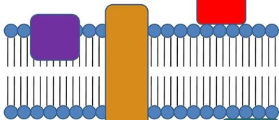
Cell Membrane: Present in all cells; also called plasma membrane, cytoplasmic membrane, or phospholipid bilayer. It defines the boundary between inside and outside the cell and is embedded with proteins.
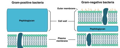
Cell Wall: Surrounds the cell membrane. Made of peptidoglycan in most bacteria, and an outer membrane in some bacteria. Distinguishes gram-positive from gram-negative bacteria.
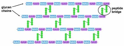
Peptidoglycan (PTG): Found only in bacteria. Provides structural rigidity and shape. Composed of long sugar chains (NAG and NAM) connected by peptide bridges. Major target of antibiotics.
Outer Membrane: Present in gram-negative bacteria. The outer leaflet is made of lipopolysaccharides (LPS), not phospholipids.
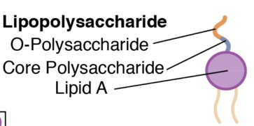
Lipopolysaccharide (LPS)
LPS is a major component of the outer membrane in gram-negative bacteria and acts as a virulence factor.
Components: O-antigen, Core polysaccharide, Lipid A (toxic part, also called endotoxin)
Function: Acts as a receptor, helps identify serotypes, and is toxic to humans. Large amounts in the bloodstream can cause septic shock and death.
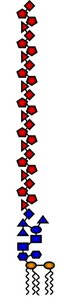
Gram Stain and Cell Envelope Types
The Gram stain divides bacteria into two major categories based on cell wall structure:
Gram-Positive: Thick peptidoglycan layer (30-50 sheets), permeable to many substances, stains purple.
Gram-Negative: Thin peptidoglycan layer (2-5 sheets), outer membrane with LPS, not permeable, stains pink.
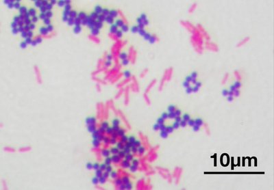
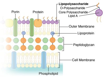
Cell Wall Exceptions
Mycoplasma spp.: No cell wall; not gram-positive or gram-negative. Causes atypical pneumonia (walking pneumonia). Antimicrobials targeting cell wall are ineffective.
Mycobacterium spp.: Extra waxy lipid layer (mycolic acid) outside peptidoglycan. Acid-fast bacteria; resist dyes, cleaning agents, and antibiotics.
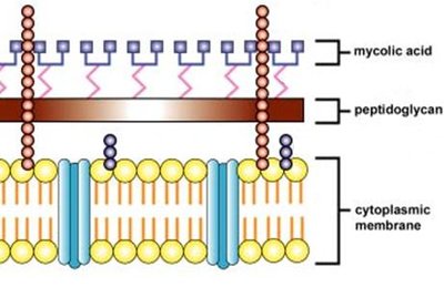
Structures Outside the Cell
Glycocalyx: Slime Layer and Capsule
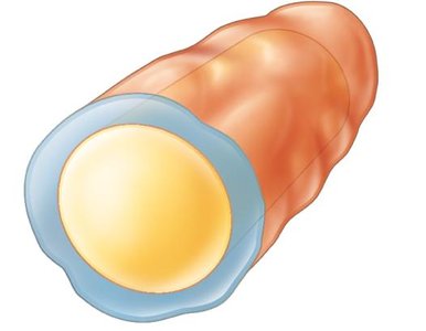
Slime Layer: Loose shield protecting bacteria from water and nutrient loss. Diffuse and irregular.
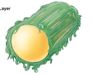
Capsule: Tightly bound, dense, thick, and gelatinous. Made of carbohydrates and/or protein. Protects from drying out, evades immune system, adheres to environment. Powerful virulence factor.
Biofilms
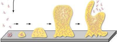
Biofilms are communities of microbes growing together on a surface, surrounded by glycocalyx.
Formation: Planktonic bacteria attach, other microbes join, glycocalyces fuse, parts break away to infect other sites.
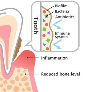
Properties: Extremely resistant to immune response, heat, chemicals, and mechanical removal. Makes microbes 1000x more resistant to antimicrobials.
Medical Relevance: Found in sewage systems, medical devices, and dental plaque.
Appendages: Flagella, Fimbriae, Pili
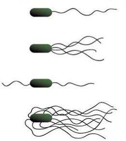
Flagella: Filamentous appendages for motility. Made of flagellin protein. Types include monotrichous (single), lophotrichous (tuft), amphitrichous (both ends), peritrichous (all over).
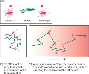
Movement: Chemotaxis (movement toward/away from chemicals), aerotaxis (oxygen), magnetotaxis (magnetic field), thermotaxis (heat), phototaxis (light). Movement consists of runs and tumbles.
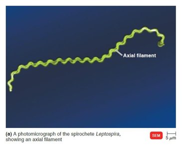
Axial Filaments: Also called endoflagella, found in spirochetes, wrapped around the cell, causing corkscrew movement.
Fimbriae: Small, bristle-like fibers for attachment to surfaces.
Pili: Hollow, rigid tubes for conjugation (DNA transfer).
Internal Structures
Cytoplasm and DNA
Cytoplasm: Gelatinous solution (cytosol) containing water, DNA, ribosomes, and other molecules.
Prokaryotic DNA: Usually circular, double-stranded, located in the nucleoid region. May also have plasmids (small, independent DNA pieces conferring traits like drug resistance).
Gene Transfer
Vertical Gene Transfer: DNA passed to next generation during binary fission.
Horizontal Gene Transfer (HGT): DNA transferred between cells of the same generation via transformation (uptake from environment), transduction (virus-mediated), and conjugation (via pilus).
Ribosomes
Function: Protein synthesis.
Structure: Made of rRNA and protein. Prokaryotic ribosomes are 70S (small subunit 30S, large subunit 50S).
Virulence Factors: Endospores and Exotoxins
Endospores
Definition: Dormant, tough, non-reproductive structures produced by some bacteria. Extremely resistant to heat, desiccation, chemicals, UV light, and boiling water.
Formation: Sporulation (vegetative cell converts to endospore under unfavorable conditions), release of free endospore, germination (endospore returns to vegetative cell in favorable conditions).
Produced by: Bacillus anthracis (anthrax), Clostridium tetani (tetanus), C. perfringens (gas gangrene), C. botulinum (botulism), Clostridioides difficile (pseudomembranous colitis).
Exotoxins
Definition: Proteins (usually enzymes) secreted by bacteria that harm the host even without the bacteria present. Major determinants of virulence.
Types: Neurotoxins (nervous system), enterotoxins (GI tract), cytotoxins (general cell function or lysis).
A-B Exotoxins: Contain an active (A) and binding (B) subunit.
Archaea
Characteristics of Archaea
Primitive Life Forms: Most closely related to the first cells on Earth.
Extremophiles: Live in extreme environments (heat, salt, acid, pH, pressure).
Evolutionary Relatedness: More closely related to Domain Eukarya than to Bacteria, based on ribosomal RNA sequences.
Summary Table: Cell Envelope Components
Component | Gram-Positive | Gram-Negative |
|---|---|---|
Cell Membrane | Present | Present |
Peptidoglycan | Thick (30-50 sheets) | Thin (2-5 sheets) |
Outer Membrane | Absent | Present (with LPS) |
LPS | Absent | Present |
Additional info: Academic context was added to clarify definitions, mechanisms, and clinical relevance for each structure and virulence factor.
