 뒤로
General Microbiology: Foundations, Cell Structure, and Microbial Diversity
스터디 가이드 - 스마트 노트
 자료에 맞춘 맞춤형 노트, 핵심 정의, 예시, 맥락을 확장해 제공합니다.
The Microbial World and You
What is Science?
Science is a systematic approach to understanding the natural world through observation, hypothesis formation, experimentation, and theory development. The scientific method is fundamental to microbiology, enabling researchers to investigate microorganisms and their effects.
Observation: Noticing phenomena in the natural world.
Hypothesis: Tentative explanation consistent with observations.
Experimentation: Testing predictions derived from the hypothesis.
Theory: Framework explaining observations and making predictions after repeated validation.
Law: Statement describing consistent natural phenomena.
Ockham’s Razor: Principle favoring simpler explanations when multiple hypotheses exist.
What is Microbiology?
Microbiology is the study of microscopic organisms, including bacteria, viruses, fungi, protozoa, and algae. It encompasses their physiology, genetics, ecology, and roles in health and disease.
Branches: Bacteriology, Virology, Mycology, Parasitology, Phycology.
Applications: Medicine, biotechnology, environmental science.
History of Microbiology
The history of microbiology is marked by key discoveries that shaped our understanding of microorganisms and their impact on health and society.
Early mentions: Microbial concepts appear in ancient texts, including the Bible.
Science begins: The formal study of microbiology began in the 17th century with the invention of microscopes.
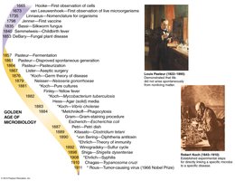
Milestones in Microbiology
Major milestones include the first observation of cells, development of vaccines, and the establishment of the germ theory of disease.
1665: Hooke observes cells.
1673: van Leeuwenhoek observes microorganisms.
1796: Jenner develops the first vaccine.
1857-1911: Golden Age of Microbiology, with discoveries by Pasteur and Koch.
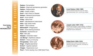
Disproving Spontaneous Generation
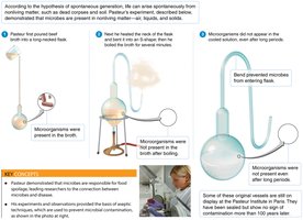
Spontaneous generation was the belief that life could arise from nonliving matter. Louis Pasteur's experiments disproved this theory, demonstrating that microorganisms originate from other microorganisms.
Pasteur's experiment: Used swan-neck flasks to show that broth remained sterile unless exposed to microbes from the air.
Controls: Included boiling, bending necks, and monitoring microbial growth.
Koch’s Postulates
Koch’s postulates are a set of criteria used to establish a causal relationship between a microorganism and a disease.
The same pathogen must be present in every case of the disease.
The pathogen must be isolated and grown in pure culture.
The cultured pathogen must cause disease in a healthy host.
The pathogen must be re-isolated and shown to be the original organism.
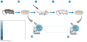
Exceptions: Some pathogens, such as Treponema pallidum (syphilis) and HIV (AIDS), do not fulfill all postulates due to difficulties in culturing or ethical constraints.
Classification of Microorganisms
Three-Domain System
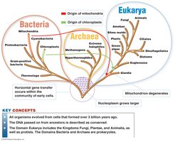
Microorganisms are classified into three domains: Bacteria, Archaea, and Eukarya. This system reflects evolutionary relationships and cellular characteristics.
Bacteria: Prokaryotic, peptidoglycan cell walls.
Archaea: Prokaryotic, no peptidoglycan, unique membrane lipids.
Eukarya: Eukaryotic, includes fungi, protozoa, algae, plants, and animals.
Cell Size Comparison
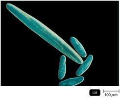
Microbial cells vary greatly in size. Eukaryotic cells are typically larger than prokaryotic cells.
Eukaryotes: 10–100 µm
Prokaryotes: 0.2–2 µm
Viruses: 0.02–0.3 µm (20–300 nm)
Volume Comparison: Assuming cubic shapes, a eukaryotic cell (20 µm) is times larger in volume than a prokaryotic cell (2 µm).
Functional Anatomy of Prokaryotic and Eukaryotic Cells
Eukaryotic Cell Structure
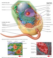
Eukaryotic cells contain membrane-bound organelles, including the nucleus, mitochondria, and chloroplasts. Plant and animal cells share many features but differ in some organelles.
Nucleus: Contains genetic material.
Mitochondria: Site of cellular respiration.
Chloroplasts: Site of photosynthesis (plants and algae).
Vacuole: Storage and structural support (plants).
Prokaryotic Cell Structure
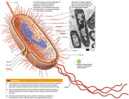
Prokaryotic cells lack membrane-bound organelles and have a simpler structure. Key components include the nucleoid, ribosomes, plasma membrane, and cell wall.
Nucleoid: Region containing DNA, usually circular and haploid.
Ribosomes: 70S type, site of protein synthesis.
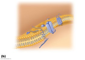
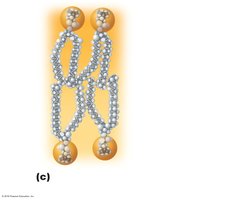
Plasma membrane: Barrier composed of proteins and lipids.
Cell wall: Provides shape and prevents osmotic lysis.
Flagella: Used for motility.
Pili and fimbriae: Used for conjugation and attachment.
Capsule: Extracellular polysaccharide for attachment.
Plasmid: Extrachromosomal DNA.
Endosymbiont Theory
The endosymbiont theory explains the origin of mitochondria and chloroplasts in eukaryotic cells, proposing that these organelles originated from symbiotic bacteria.
Mitochondria: Derived from aerobic bacteria.
Chloroplasts: Derived from photosynthetic bacteria.
Evidence: Both organelles contain their own DNA and resemble prokaryotes.
Shapes and Arrangements of Prokaryotic Cells
Bacteria exhibit diverse shapes and arrangements, which are important for identification and classification.
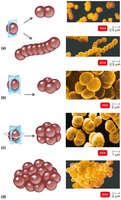
Cocci: Spherical, can form diplococci, streptococci, tetrads, sarcinae, staphylococci.
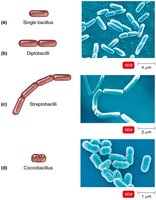
Bacilli: Rod-shaped, can be single, diplobacilli, streptobacilli, coccobacilli.
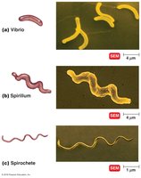
Spiral: Includes vibrio, spirillum, spirochete.
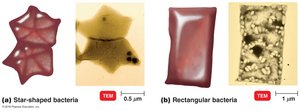
Other shapes: Star-shaped, rectangular.
Prokaryotic Cell Wall and Gram Stain
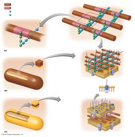
The cell wall is a critical structure in prokaryotes, providing shape and protection. The Gram stain differentiates bacteria based on cell wall composition.
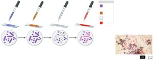
Gram-positive: Thick peptidoglycan layer, retains crystal violet stain (purple).
Gram-negative: Thin peptidoglycan, outer membrane with lipopolysaccharide (LPS), loses crystal violet, stains red with safranin.
Gram Stain Steps: Crystal violet, iodine, alcohol decolorization, safranin counterstain.
Osmosis and Cell Wall Function
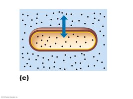
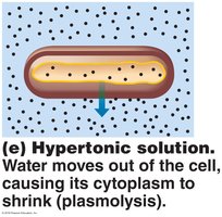
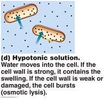
The cell wall prevents osmotic lysis and maintains cell shape. Osmosis is the movement of water across membranes, affecting cell integrity.
Isotonic: No net water movement.
Hypertonic: Water leaves cell, causing plasmolysis.
Hypotonic: Water enters cell, causing swelling or lysis if wall is weak.
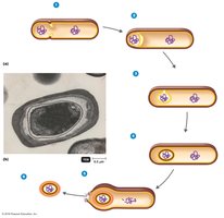
Endospores
Endospores are highly resistant structures formed by certain bacteria, allowing survival in extreme conditions.
Formation: Sporulation process produces endospores.
Resistance: High temperature, toxic chemicals, desiccation, radiation, low temperatures.
Diseases: Bacillus anthracis (anthrax), Clostridium perfringens (gas gangrene), C. tetani (tetanus), C. botulinum (botulism).
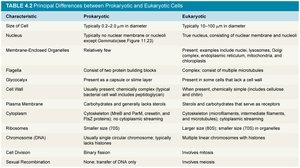
Principal Differences between Prokaryotic and Eukaryotic Cells
Prokaryotic and eukaryotic cells differ in structure, genetic material, and cellular processes.
Characteristic | Prokaryotic | Eukaryotic |
|---|---|---|
Size of Cell | 0.2–2 µm | 10–100 µm |
Nucleus | No nuclear membrane | True nucleus with nuclear membrane |
Membrane-Enclosed Organelles | Absent | Present (mitochondria, ER, etc.) |
Flagella | Composed of protein | Complex, microtubules |
Cell Wall | Peptidoglycan (bacteria) | Cellulose (plants), chitin (fungi) |
Ribosomes | 70S | 80S |
Chromosome (DNA) | Single, circular | Multiple, linear |
Cell Division | Binary fission | Mitosis |
The Eukaryotes: Fungi, Algae, Protozoa
Fungi
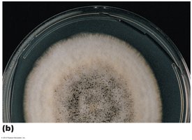
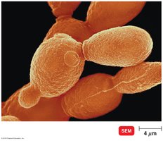
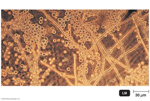
Fungi are eukaryotic organisms that include molds and yeasts. They play important roles in disease, food production, and biotechnology.
Molds: Multicellular, aerobic, form hyphae.
Yeasts: Single-celled, facultative anaerobes, reproduce by budding.
Dimorphism: Some fungi can exist as both molds and yeasts.
Importance: Diseases, food (cheese, bread), antibiotics (penicillin), drugs.
Protozoa
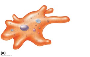
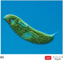
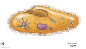
Protozoa are single-celled eukaryotes classified by their motility structures. They are important in the food chain and as disease agents.
Pseudopodia: Amoeboid movement (e.g., Amoeba).
Flagella: Whip-like movement (e.g., Euglena).
Cilia: Hair-like movement (e.g., Paramecium).
Cytoplasm types: Ectoplasm (outer), endoplasm (inner).
Diseases: Malaria, amoebic dysentery.
Algae
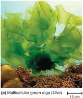
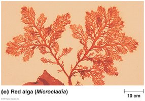
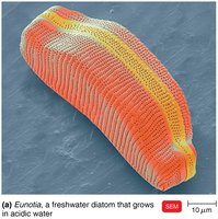
Algae are photosynthetic eukaryotes with chloroplasts. They are important for oxygen production and as sources of agar. Some algae cause diseases, such as Paralytic Shellfish Poisoning.
Green algae: Ulva, multicellular.
Red algae: Microcladia, used in agar production.
Diatoms: Silica cell walls, important in aquatic ecosystems.
Additional info:
Some details, such as the full scope of Ockham’s Razor or the specifics of certain atypical bacteria, were inferred for completeness.
Tables and diagrams were recreated and expanded for clarity and academic context.
