 뒤로
뒤로Genetics: Structure, Function, and Variation of Genetic Material
스터디 가이드 - 스마트 노트
 자료에 맞춘 맞춤형 노트, 핵심 정의, 예시, 맥락을 확장해 제공합니다.
Genetics: Structure, Function, and Variation of Genetic Material
Introduction to Genetics
Genetics is the study of genes, their functions, and how variations arise in genomes. The genome is the entire collection of genetic material in a cell or virus. A gene is a heritable unit of genetic material that determines a particular trait. The genotype refers to the genetic makeup of an organism, while the phenotype is the observable collection of traits.
Genotype: The inherited set of genetic instructions contained in the genome.
Phenotype: The organism’s observable traits (physical and physiological characteristics).
Organization of Genetic Material
Prokaryotic vs. Eukaryotic Cells
Genetic material is organized differently in prokaryotic and eukaryotic cells.
Eukaryotic cells:
Numerous linear chromosomes housed in the nucleus
Organizational proteins called histones prevent DNA tangling
Genetic material also found in mitochondria and chloroplasts
Prokaryotic cells:
1–3 chromosomes, usually circular, located in the nucleoid region
Histone-like proteins organize DNA
Contain chromosomal DNA and plasmids
Plasmids
Plasmids are small, circular DNA molecules that exist outside the chromosomal DNA in bacteria and some eukaryotic cells. They often carry genes conferring antibiotic resistance.
Structure of DNA and RNA
Nucleotide Structure
Both DNA and RNA are polymers of nucleotides, each consisting of three components:
Phosphate group
Sugar (deoxyribose in DNA, ribose in RNA)
Nitrogenous base (Adenine, Guanine, Cytosine, Thymine in DNA; Uracil replaces Thymine in RNA)
Nitrogenous Bases: Classification and Pairing
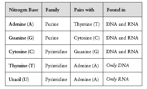
Nitrogenous bases are classified as purines (double-ring) or pyrimidines (single-ring). Base pairing is specific: A pairs with T (or U in RNA), and G pairs with C.
Nitrogen Base | Family | Pairs with | Found in |
|---|---|---|---|
Adenine (A) | Purine | Thymine (T) | DNA and RNA |
Guanine (G) | Purine | Cytosine (C) | DNA and RNA |
Cytosine (C) | Pyrimidine | Guanine (G) | DNA and RNA |
Thymine (T) | Pyrimidine | Adenine (A) | Only DNA |
Uracil (U) | Pyrimidine | Adenine (A) | Only RNA |
DNA Structure
Double-stranded molecule forming a double helix (twisted ladder)
“Rungs” are complementary nitrogen bases (A–T, G–C)
“Side rails” are alternating sugar and phosphate groups, linked by phosphodiester bonds
Antiparallel orientation: one strand runs 5’→3’, the other 3’→5’
RNA Structure
Usually single-stranded
Contains ribose sugar
Uracil (U) replaces thymine (T)
Can fold into helical and loop structures
Central Dogma of Molecular Biology
Flow of Genetic Information
The central dogma describes the flow of genetic information: DNA → RNA → Protein.
Transcription: DNA is used as a template to synthesize RNA.
Translation: RNA (specifically mRNA) is decoded by ribosomes to build proteins.
DNA Replication
Overview and Enzymes
DNA replication is the process by which a cell copies its genome before division. It is semiconservative: each new DNA molecule contains one original and one new strand.
Helicase: Unwinds the DNA helix.
Gyrase/Topoisomerase: Relieves supercoiling ahead of the replication fork.
Primase: Synthesizes short RNA primers to initiate DNA synthesis.
DNA Polymerase III: Main enzyme that adds nucleotides in the 5’→3’ direction.
DNA Polymerase I: Removes RNA primers and replaces them with DNA.
Ligase: Seals nicks in the sugar-phosphate backbone, joining Okazaki fragments.
Single-stranded binding proteins: Stabilize separated DNA strands.
Replication proceeds differently on the two strands:
Leading strand: Synthesized continuously toward the replication fork.
Lagging strand: Synthesized discontinuously away from the fork in Okazaki fragments.
Transcription and Translation
Transcription
RNA polymerase binds to the promoter region of DNA.
Unwinds DNA and synthesizes complementary RNA (A pairs with U in RNA).
Stops at a termination sequence; RNA transcript is released.
Types of RNA produced:
mRNA (messenger RNA): Carries genetic instructions to ribosomes.
tRNA (transfer RNA): Brings amino acids to ribosomes; contains an anticodon loop.
rRNA (ribosomal RNA): Forms part of the ribosome and catalyzes peptide bond formation.
Translation
Ribosomes read mRNA codons (triplets of nucleotides).
Each codon specifies an amino acid or a stop signal.
tRNA molecules bring the correct amino acids, matching codons with anticodons.
Ribosomes catalyze peptide bond formation, building the protein.
The genetic code is redundant: multiple codons can specify the same amino acid.
Gene Regulation
Pre-Transcriptional Regulation
Operons (in prokaryotes): clusters of genes regulated together
Epigenetic control (e.g., DNA methylation)
Recruitment of transcription factors
Post-Transcriptional Regulation
Control of mRNA stability and translation efficiency
Small noncoding RNAs and riboswitches
RNA processing (e.g., splicing in eukaryotes)
Mutations and Genetic Variation
Types of Mutations
Silent: No change in amino acid sequence
Missense: Changes one amino acid
Nonsense: Changes codon to a stop signal
Frameshift: Insertion or deletion shifts reading frame
Mutations can be spontaneous (natural errors) or induced (by mutagens such as chemicals or radiation).
DNA Repair Mechanisms
Proofreading: DNA polymerases correct errors during replication.
Excision repair: Damaged or mismatched nucleotides are removed and replaced.
Horizontal Gene Transfer in Bacteria
Conjugation: DNA transfer via direct cell contact and a pilus.
Transformation: Uptake of free DNA from the environment.
Transduction: Transfer of DNA by bacteriophages (viruses).
Transposons
"Jumping genes" that can move within the genome
Retrotransposons use reverse transcriptase to copy RNA into DNA
DNA transposons move via "cut-and-paste" or "copy-and-paste" mechanisms
Summary Table: Nitrogenous Bases
Nitrogen Base | Family | Pairs with | Found in |
|---|---|---|---|
Adenine (A) | Purine | Thymine (T) | DNA and RNA |
Guanine (G) | Purine | Cytosine (C) | DNA and RNA |
Cytosine (C) | Pyrimidine | Guanine (G) | DNA and RNA |
Thymine (T) | Pyrimidine | Adenine (A) | Only DNA |
Uracil (U) | Pyrimidine | Adenine (A) | Only RNA |
Key Equations and Concepts
Phosphodiester bond formation:
Central Dogma:
Genetic code: 64 codons (triplets of nucleotides) encode 20 amino acids, 1 start signal, and 3 stop signals.
Examples and Applications
Example of a silent mutation: GAA (glutamate) → GAG (glutamate)
Example of a missense mutation: GAA (glutamate) → GUA (valine)
Example of a nonsense mutation: GAA (glutamate) → UAA (stop codon)
Example of frameshift mutation: Inserting or deleting a base shifts the reading frame, altering downstream amino acids.
Additional info: The provided table and image reinforce the classification and pairing of nitrogenous bases, which is foundational for understanding DNA and RNA structure and function.
