 뒤로
Gram-Positive Bacterial Diseases of the Skin and Related Systems
스터디 가이드 - 스마트 노트
 자료에 맞춘 맞춤형 노트, 핵심 정의, 예시, 맥락을 확장해 제공합니다.
Bacterial Diseases: Gram Positive Organisms
Overview of Gram-Positive Bacterial Infections
Gram-positive bacteria are responsible for a wide range of skin and systemic infections. These organisms include Mycobacterium tuberculosis, Bacillus anthracis, Clostridium species, Staphylococcus species, Streptococcus species, and Propionibacterium acnes. Their pathogenicity is often linked to unique structural and biochemical features, as well as their ability to evade host defenses.
Mycobacterium tuberculosis and Tuberculosis
Pathogenesis and Clinical Features
Mycobacterium tuberculosis is an acid-fast, high G+C, gram-positive rod with a waxy cell wall rich in mycolic acids. This structure allows the bacterium to resist phagocytosis and survive within macrophages, leading to chronic granulomatous disease, primarily affecting the lungs.
Transmission: Inhalation of respiratory droplets or aerosols.
Infectious Dose: As few as 10 cells.
Immune Response: Formation of tubercles (walled-off lesions) in alveoli; chronic inflammation and tissue damage.
Progression: Tubercles may rupture, leading to miliary tuberculosis (systemic spread).
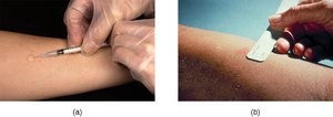
Diagnosis: Chest radiographs (Ghon complexes), Mantoux tuberculin skin test.
Treatment: Multidrug protocols (isoniazid, rifampin, ethambutol, pyrazinamide).
Prevention: BCG vaccine (limited use in the US).
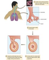
Example: Formation of tubercles in the lung is a hallmark of TB, visible in radiographs as Ghon complexes.
Bacillus anthracis and Anthrax
Types and Pathogenesis
Bacillus anthracis is a gram-positive, endospore-forming facultative anaerobe. Anthrax can be cutaneous, gastrointestinal, pulmonary, or injectional, with cutaneous anthrax being the most common.
Cutaneous Anthrax: Entry through skin abrasions; formation of black eschar.
Virulence Factors: Capsule prevents phagocytosis; binary exotoxins cause edema and tissue damage.
Diagnosis: Blood agar culture, clinical presentation.
Treatment: Broad-spectrum antibiotics (penicillin, erythromycin, tetracycline).
Prevention: Vaccine for at-risk individuals.
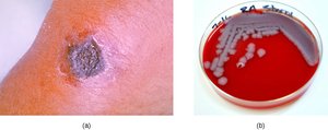
Example: Cutaneous anthrax presents as a black eschar, a mass of dead skin tissue.
Clostridium-Associated Diseases
Tetanus and Botulism
Species of Clostridium are gram-positive, endospore-forming rods and obligate anaerobes. They produce potent exotoxins, including tetanus neurotoxin (TeNT) and botulinum neurotoxin (BoNT).
Tetanus: Caused by C. tetani; characterized by uncontrollable muscle spasms due to TeNT blocking GABA release.
Botulism: Caused by C. botulinum; BoNT blocks acetylcholine release, leading to flaccid paralysis.
Diagnosis: Bioassays, ELISA, RT-PCR.
Treatment: Antitoxins, assisted breathing, antibiotics (for tetanus).
Prevention: Tetanus toxoid vaccine (DTaP, Tdap, Td).
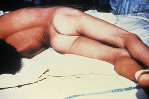
Example: Opisthotonos is a rigid, arched posture seen in severe tetanus cases.
Clostridium difficile Infections
Pathogenesis and Treatment
Clostridium difficile can cause antibiotic-associated diarrhea and pseudomembranous colitis, especially in immunocompromised patients or those with disrupted gut microbiota.
Toxins: TcdA and TcdB inactivate GTP-binding proteins, causing cell death and inflammation.
Diagnosis: Detection of toxins in stool, PCR, ELISA.
Treatment: Stop antibiotics, supportive therapy, metronidazole or vancomycin, fecal transplant.
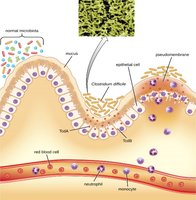
Example: Pseudomembranous colitis is characterized by the formation of a pseudomembrane in the colon.
Staphylococcus Species
Pathogenicity and Diagnosis
Staphylococcus aureus is a common cause of skin and systemic infections, often purulent due to leukocidin production. Methicillin-resistant S. aureus (MRSA) is a major concern in healthcare settings.
Virulence Factors: Hemolysins, coagulase, leukocidins, Protein A, enterotoxins.
Diagnosis: Gram stain (grapelike clusters), catalase test (positive), coagulase test, mannitol salt agar (MSA).
Treatment: Antibiotic sensitivity testing, MRSA-effective drugs.
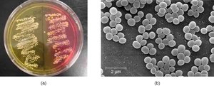
Example: S. aureus ferments mannitol on MSA, turning the medium yellow.
Superficial Staphylococcal Infections
Folliculitis: Localized rash due to inflammation of hair follicles.
Furuncles (boils): Deeper, pus-filled abscesses.
Carbuncles: Deep, pus-filled abscesses formed from multiple furuncles.
Staphylococcal Scalded Skin Syndrome (SSSS): Exotoxins cause erythema and severe skin peeling.
Impetigo: Vesicles, pustules, bullae; highly contagious, often in children.
Example: Furuncles and carbuncles are common staphylococcal skin infections; SSSS causes severe skin peeling.
Staphylococcal Food Poisoning
Enterotoxins: Cause rapid onset of nausea, vomiting, diarrhea.
Diagnosis: Identification of toxin in food or biological specimens.
Prevention: Proper food handling, cooking, and storage.
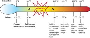
Example: Safe internal temperatures for food storage and cooking prevent staphylococcal food poisoning.
Streptococcus Species
Pathogenicity and Diagnosis
Streptococcus pyogenes (Group A Streptococcus, GAS) is a major pathogen, producing extracellular enzymes and virulence factors such as streptolysins, hyaluronidase, streptokinase, capsule, and M protein.
Microscopy: Chains of cocci, catalase-negative.
Diagnosis: Agglutination reactions, ELISA, bacitracin susceptibility.
Treatment: Penicillin (resistance not widespread).
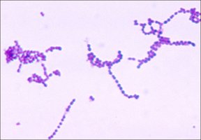
Example: Streptococcus pyogenes forms chains of cocci under the microscope.
Streptococcal Skin Conditions
Cellulitis: Red, warm, painful area of skin.
Erysipelas: Large, intensely inflamed patch of skin.
Erythema Nodosum: Red nodules, usually on the shins.
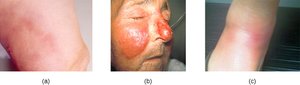
Example: Cellulitis, erysipelas, and erythema nodosum are common streptococcal skin conditions.
Necrotizing Fasciitis
Necrotizing fasciitis is a rare but life-threatening condition caused by S. pyogenes and other bacteria. It involves rapid destruction of fascia and surrounding tissues, often requiring surgical debridement or amputation.
Virulence Factors: Adhesion, invasion, proteases, complement inactivation.
Treatment: Surgical removal, intravenous antibiotics.
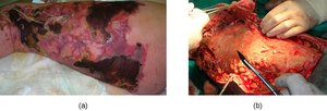
Example: Necrotizing fasciitis requires aggressive surgical and medical intervention.
Propionibacterium acnes and Acne
Pathogenesis and Treatment
Propionibacterium acnes is a gram-positive, non-spore-forming, aerotolerant anaerobic bacillus found on skin. It contributes to acne by consuming sebum and secreting enzymes that damage hair follicles, leading to inflammatory lesions.
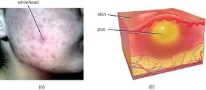
Lesion Types: Comedones (whiteheads, blackheads), papules, pustules, nodules, pseudocysts.
Treatment: Topical agents (salicylic acid, retinoids), antibiotics, hormone therapy, isotretinoin, phototherapy.
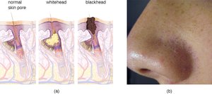
Example: Acne is characterized by whitehead and blackhead comedones; severe cases may require advanced therapies.
Medical Terms Associated with Skin Lesions and Rashes
Definitions
Term | Definition |
|---|---|
Abscess | Localized collection of pus |
Bulla | Fluid-filled blister >5 mm diameter |
Carbuncle | Deep, pus-filled abscess from multiple furuncles |
Crust | Dried fluids from a lesion |
Cyst | Encapsulated sac filled with fluid, semi-solid matter, or gas |
Folliculitis | Localized rash due to inflammation of hair follicles |
Furuncle | Pus-filled abscess due to infection of a hair follicle |
Macules | Smooth spots of discoloration |
Papules | Small raised bumps |
Pseudocyst | Lesion resembling a cyst but less defined |
Purulent | Pus-producing; suppurative |
Pustules | Fluid- or pus-filled bumps |
Pyoderma | Suppurative infection of the skin |
Suppurative | Producing pus; purulent |
Ulcer | Break in the skin; open sore |
Vesicle | Small, fluid-filled lesion |
Wheal | Swollen, inflamed skin that itches or burns |
Example: Understanding these terms is essential for describing and diagnosing skin lesions.
Summary Table: Comparison of Gram-Positive Skin Pathogens
Pathogen | Key Features | Common Diseases | Diagnosis | Treatment |
|---|---|---|---|---|
Mycobacterium tuberculosis | Acid-fast, waxy cell wall | Tuberculosis | Radiographs, skin test | Multidrug therapy |
Bacillus anthracis | Endospore-forming | Anthrax | Blood agar, clinical | Antibiotics, vaccine |
Clostridium spp. | Endospore-forming, exotoxins | Tetanus, botulism, gastroenteritis | Bioassays, ELISA | Antitoxins, antibiotics |
Staphylococcus aureus | Coagulase-positive, clusters | Pyoderma, SSSS, impetigo, food poisoning | Culture, MSA, coagulase test | Antibiotics, hygiene |
Streptococcus pyogenes | Chains, catalase-negative | Cellulitis, erysipelas, necrotizing fasciitis | Agglutination, ELISA | Penicillin, surgery |
Propionibacterium acnes | Non-spore-forming, anaerobic | Acne | Clinical, visual | Topical, antibiotics |
