 뒤로
Gram Stain: Principles, Procedure, and Interpretation
스터디 가이드 - 스마트 노트
 자료에 맞춘 맞춤형 노트, 핵심 정의, 예시, 맥락을 확장해 제공합니다.
Gram Stain
Introduction to the Gram Stain
The Gram stain is a fundamental differential staining technique in microbiology used to classify bacteria into two major groups: Gram-positive and Gram-negative. This distinction is based on differences in the structure and composition of bacterial cell walls, which affect their ability to retain certain dyes during the staining process. The Gram stain is essential for bacterial identification, guiding treatment decisions, and understanding microbial physiology.
Cell Wall Structure and Its Role in Gram Staining
Gram-Positive vs. Gram-Negative Cell Walls
Bacterial cell walls are the primary determinant of the Gram reaction. The differences in cell wall structure explain why Gram-positive and Gram-negative bacteria stain differently.
Gram-Positive Bacteria: Thick peptidoglycan layer, presence of teichoic acids, no outer membrane.
Gram-Negative Bacteria: Thin peptidoglycan layer, outer membrane containing lipopolysaccharides, presence of porins.
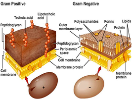
Example: Staphylococcus epidermidis is Gram-positive; Escherichia coli is Gram-negative.
Principles of the Gram Stain
How the Gram Stain Works
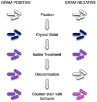
The Gram stain involves a series of steps using different reagents. The outcome depends on the cell wall's ability to retain or lose the primary dye after decolorization.
Primary Stain (Crystal Violet): Stains all cells purple.
Mordant (Iodine): Forms a complex with crystal violet, trapping it in the cell wall.
Decolorization (Alcohol or Acetone): Removes the dye from Gram-negative cells but not from Gram-positive cells.
Secondary Stain (Safranin): Counterstains decolorized cells pink/red.
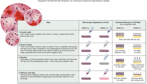
Key Point: The thick peptidoglycan layer in Gram-positive bacteria retains the crystal violet-iodine complex, while the thin layer in Gram-negative bacteria does not.
Gram Stain Procedure
Step-by-Step Protocol
The Gram stain is performed in a specific sequence to ensure accurate differentiation:
Application of crystal violet (1 minute)
Application of iodine (1 minute)
Alcohol wash (generally no more than 10 seconds; cell density dependent)
Application of safranin (1 minute)
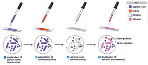
Additional info: Over-decolorization can cause Gram-positive cells to appear Gram-negative; under-decolorization can cause Gram-negative cells to appear Gram-positive.
Microscopic Appearance and Interpretation
Observing Gram Stained Slides
After staining, Gram-positive bacteria appear purple, and Gram-negative bacteria appear pink/red under the microscope. The shape and arrangement of cells can also be observed, aiding in identification.
Gram-Positive: Typically cocci (spherical) in clusters or chains.
Gram-Negative: Typically rods (bacilli) or other shapes.
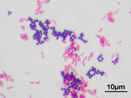
Example: In a mixed culture, S. epidermidis (purple cocci) and E. coli (pink rods) can be distinguished.
Critical Steps and Troubleshooting
Tips for a Successful Gram Stain
Decolorization is crucial: Too much or too little can lead to incorrect results.
Good emulsion: Proper bacterial smear preparation ensures even staining.
Gram variability: Some Gram-positive bacteria (e.g., Bacillus species) may appear Gram-variable in older cultures (>24 hours).
Result of over-decolorization: Gram-positive cells may appear pink (false Gram-negative).
Result of under-decolorization: Gram-negative cells may appear purple (false Gram-positive).
Summary Table: Gram Stain Steps and Results
Step | Gram-Positive Appearance | Gram-Negative Appearance |
|---|---|---|
Crystal Violet | Purple | Purple |
Iodine Treatment | Purple | Purple |
Decolorization | Purple | Colorless |
Counterstain (Safranin) | Purple | Pink/Red |
Applications and Laboratory Practice
Practical Use of the Gram Stain
In the laboratory, the Gram stain is used to analyze pure and mixed cultures, as well as clinical samples (e.g., gumline swabs). Proper disposal of slides and adherence to safety protocols are essential. The Gram stain guides further testing and initial identification of bacterial pathogens.
Example: Slides prepared from E. coli, S. epidermidis, and gumline samples are stained and examined to determine Gram reaction and morphology.
