 뒤로
Gram Staining: Principles, Procedure, and Interpretation
스터디 가이드 - 스마트 노트
 자료에 맞춘 맞춤형 노트, 핵심 정의, 예시, 맥락을 확장해 제공합니다.
Gram Staining
Introduction to Gram Staining
The Gram stain is a fundamental differential staining technique in microbiology, developed by Hans Christian Gram in 1884. It is used to classify bacteria into two major groups—Gram-positive and Gram-negative—based on differences in their cell wall structure. This distinction is crucial for bacterial identification, understanding pathogenicity, and guiding antibiotic therapy.
Principle of the Gram Stain
Gram-positive bacteria have a thick peptidoglycan layer in their cell wall, which retains the primary stain and appears purple under the microscope.
Gram-negative bacteria possess a thinner peptidoglycan layer and an outer membrane containing lipopolysaccharides. They do not retain the primary stain after decolorization and instead take up the counterstain, appearing pink.
The Gram stain is most reliable on young, actively growing bacterial cultures.
Gram Stain Procedure
The Gram stain involves a series of steps, each with a specific purpose:
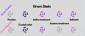
Fixation: Bacterial smears are fixed to the slide by heat to preserve cell morphology.
Primary Stain (Crystal Violet): All cells are stained purple.
Mordant (Gram's Iodine): Iodine binds with crystal violet, forming a complex that is more difficult to remove from Gram-positive cells.
Decolorization (95% Ethanol): Alcohol dissolves the outer membrane of Gram-negative cells and removes the crystal violet-iodine complex, leaving them colorless. Gram-positive cells retain the purple color.
Counterstain (Safranin): Stains decolorized Gram-negative cells pink, while Gram-positive cells remain purple.
Examination: Slides are observed under oil immersion (100x objective) for cell morphology and color.
Step-by-Step Protocol
Prepare and fix bacterial smears on clean slides.
Stain with crystal violet for 60 seconds, rinse with distilled water, and blot dry.
Add Gram's iodine for 30 seconds, rinse with water, and blot dry.
Decolorize with 95% ethanol until runoff is clear (about 10 seconds), then blot dry.
Counterstain with safranin for 60 seconds, rinse with water, and blot dry.
Examine under the microscope, progressing from low to oil immersion magnification.
Interpretation of Results
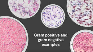
Gram-positive cells: Appear purple due to retention of the crystal violet-iodine complex. Example: Staphylococcus epidermidis, Bacillus subtilis.
Gram-negative cells: Appear pink due to uptake of safranin. Example: Escherichia coli.
Cell Wall Structure and Antibiotic Sensitivity
Gram-positive bacteria: Thick peptidoglycan layer; generally more susceptible to antibiotics like penicillin and cephalosporins.
Gram-negative bacteria: Outer membrane with lipopolysaccharides; often more resistant to antibiotics due to this barrier.
Limitations of the Gram Stain
Best results are obtained with young, actively growing cultures.
Some bacteria may not stain reliably due to cell wall abnormalities or age.
Summary Table: Gram Stain Steps and Outcomes
Step | Reagent | Purpose | Gram-Positive Appearance | Gram-Negative Appearance |
|---|---|---|---|---|
Fixation | Heat | Adheres cells to slide | Colorless | Colorless |
Primary Stain | Crystal Violet | Stains all cells purple | Purple | Purple |
Mordant | Gram's Iodine | Forms complex with crystal violet | Purple | Purple |
Decolorization | 95% Ethanol | Removes stain from Gram-negative cells | Purple | Colorless |
Counterstain | Safranin | Stains Gram-negative cells pink | Purple | Pink |
Examples and Applications
Clinical diagnosis: Rapid identification of bacterial pathogens in patient samples.
Antibiotic selection: Guides initial therapy based on Gram reaction.
Research: Differentiation of bacterial isolates in laboratory studies.
Practice Questions
What color will a Gram-negative cell stain after the procedure?
What color will a Gram-positive cell stain after the procedure?
Which organism used in this exercise is a coccus?
Of the three bacteria studied, which is the largest?
Additional info: The Gram stain is a foundational technique in microbiology labs and is essential for both clinical and research applications. Mastery of the procedure and interpretation is critical for accurate bacterial identification and understanding of microbial physiology.
