 뒤로
Introduction to Microbiology: Foundations, Classification, and Laboratory Techniques
스터디 가이드 - 스마트 노트
 자료에 맞춘 맞춤형 노트, 핵심 정의, 예시, 맥락을 확장해 제공합니다.
Introduction to Microbiology
Overview of Microbiology
Microbiology is the scientific study of microbes, which include a diverse group of organisms and nonliving entities. Understanding microbes is essential for comprehending human health, disease, and the ecological roles of microorganisms.
Microbes: Encompass bacteria, archaea, fungi, protists, helminths, viruses, and prions.
Humans coexist with hundreds of bacterial species, forming complex ecosystems.
Most microbes are not pathogenic; only a minority cause disease.
Major Groups of Microbes
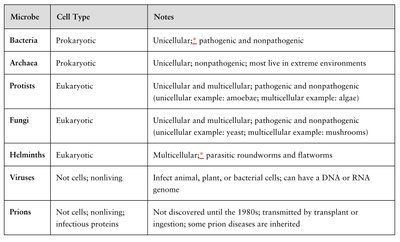
Microbes are classified based on cell type, structure, and function. The table below summarizes the main groups:
Microbe | Cell Type | Notes |
|---|---|---|
Bacteria | Prokaryotic | Unicellular; pathogenic and nonpathogenic |
Archaea | Prokaryotic | Unicellular; nonpathogenic; most live in extreme environments |
Protists | Eukaryotic | Unicellular and multicellular; pathogenic and nonpathogenic (e.g., amoebae, algae) |
Fungi | Eukaryotic | Unicellular and multicellular; pathogenic and nonpathogenic (e.g., yeast, mushrooms) |
Helminths | Eukaryotic | Multicellular; parasitic roundworms and flatworms |
Viruses | Not cells; nonliving | Infect animal, plant, or bacterial cells; can have DNA or RNA genome |
Prions | Not cells; nonliving infectious proteins | Discovered in the 1980s; transmitted by transplant or ingestion; some are inherited |
A Brief History of Microbiology
Key Discoveries and Theories
The field of microbiology has evolved through pivotal discoveries and the development of foundational theories:
Robert Hooke: First descriptions of cells using a microscope.
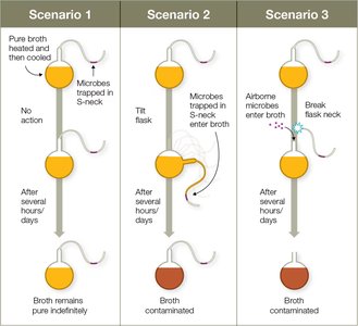
Spontaneous Generation vs. Biogenesis: Early debates on whether life arises from nonliving matter (spontaneous generation) or from existing life (biogenesis).
Louis Pasteur: Disproved spontaneous generation through experiments demonstrating that microbes do not arise from nonliving broth unless contaminated. Developed pasteurization to reduce pathogens in food.
Germ Theory of Disease: Microbes are causative agents of infectious diseases.
Koch’s Postulates: Criteria to establish a microbe as the cause of a disease:
Same organism present in every case of the disease.
Organism isolated and grown in pure culture.
Isolated organism causes disease in a susceptible host.
Organism re-isolated from the experimentally infected host.
Aseptic Techniques: Procedures to prevent contamination, pioneered by Ignaz Semmelweis and Joseph Lister (e.g., handwashing, sterilization).
Scientific Method and Terminology
Observation: Describing phenomena.
Conclusion: Inferring explanations based on observations.
Law: Predicts what happens (e.g., gravity).
Theory: Explains how or why something happens, supported by extensive evidence (e.g., cell theory).
Classifying Microbes and Their Interactions
Taxonomy and Nomenclature
Taxonomy is the science of classifying organisms based on shared characteristics. Carl Linnaeus established the modern system of taxonomy.
Hierarchy: Domain, Kingdom, Phylum, Class, Order, Family, Genus, Species.
Three Domains: Bacteria, Archaea, Eukarya.
Binomial Nomenclature: Scientific names use genus and species (e.g., Escherichia coli).
Strain: Genetic variant within a species.
Symbiotic Relationships
Symbiosis: Close association between two or more organisms.
Parasitism: One organism benefits, the host is harmed (e.g., helminths, protozoans).
Mutualism: Both organisms benefit (e.g., humans and gut bacteria).
Commensalism: One benefits, the other is unaffected (e.g., some skin microbes).
Normal and Transient Microbiota
Normal Microbiota: Permanent residents of the human body, usually protective.
Opportunistic Pathogens: Cause disease when host defenses are compromised.
Transient Microbiota: Temporary residents acquired from the environment.
Biofilms
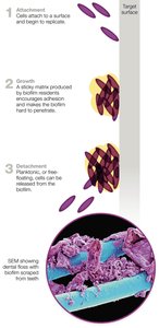
Biofilms are structured communities of microbes attached to surfaces and embedded in a self-produced matrix. They are medically significant due to their resistance to antibiotics and role in chronic infections.
Formed by single or multiple species.
Examples: Dental plaque, catheter-associated infections.
Biofilms are more resistant than planktonic (free-floating) cells.
Microbes in Human Health and Industry
Microbes aid in digestion, produce vitamins, and protect against pathogens.
Used in food production (e.g., yogurt, bread), pharmaceuticals (e.g., antibiotics), and bioremediation (cleaning up pollutants).
Growing, Staining, and Viewing Microbes
Laboratory Culturing Techniques
Growth Media: Nutrient mixtures (e.g., nutrient agar, broth) for culturing microbes.
Pure Culture: Contains only one microbial species.
Aseptic Technique: Prevents contamination during culturing.
Streak Plate Technique: Isolates individual colonies from a mixed sample.
Colony: Genetically identical group of cells from a single parent cell.
Staining Techniques
Stains: Dyes used to increase contrast and visualize microbes.
Simple Staining: Uses one dye (e.g., methylene blue).
Differential Staining: Distinguishes between cell types (e.g., Gram stain, acid-fast stain).
Gram Stain:
Gram-positive: Thick peptidoglycan, stains purple.
Gram-negative: Thin peptidoglycan, outer membrane, stains pink.
Acid-Fast Stain: Identifies bacteria with waxy cell walls (e.g., Mycobacterium).
Special Stains: Highlight structures like flagella, capsules, and endospores.
Microscopy
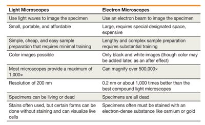
Microscopes are essential tools for visualizing microbes. There are two main types: light microscopes and electron microscopes.
Light Microscopes | Electron Microscopes |
|---|---|
Use light waves to image specimen | Use electron beam to image specimen |
Small, portable, affordable | Large, expensive, require special space |
Simple sample preparation | Complex, lengthy preparation |
Color images possible | Only black and white (color may be added later) |
Max magnification ~1,000x | Can magnify over 500,000x |
Resolution ~200 nm | Resolution ~0.2 nm |
Live or dead specimens | Only dead specimens |
Stains often used; some forms allow live cell imaging | Specimens must be stained with electron-dense substances |
Compound Light Microscope: Most common; uses objective and ocular lenses for magnification.
Bright Field Microscopy: Best for stained, non-living specimens.
Electron Microscopy:
Transmission Electron Microscope (TEM): Produces 2D images of thin sections.
Scanning Electron Microscope (SEM): Produces 3D images of surfaces.
Fluorescence Microscopy: Uses fluorescent dyes (fluorochromes) to visualize specific structures or proteins, often linked to antibodies (immunofluorescence).
Summary of Key Laboratory Techniques
Staining and microscopy are fundamental for identifying and classifying microbes.
Proper culturing and aseptic techniques are essential for accurate study and diagnosis.
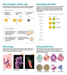
Additional info: The images included reinforce key concepts such as microbial classification, Pasteur's experiment, biofilm formation, microscopy comparisons, and laboratory techniques. All tables have been recreated in HTML for clarity and accessibility.
