 뒤로
뒤로Microbial Cell Structure and Function: Comprehensive Study Notes
스터디 가이드 - 스마트 노트
 자료에 맞춘 맞춤형 노트, 핵심 정의, 예시, 맥락을 확장해 제공합니다.
Microbial Morphology
Cell Shape and Major Morphologies
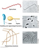
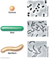
Microbial morphology refers to the shape and arrangement of microbial cells, which is a key characteristic used in identification. The three primary shapes are coccus (spherical), rod (bacillus), and spirillum (spiral). Some bacteria exhibit unusual shapes such as spirochetes, stalked, hyphal, and filamentous forms. These morphologies are genetically encoded and often optimized for specific environmental conditions.
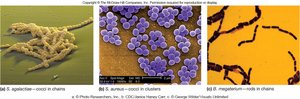
Coccus: Spherical shape, often found in clusters or chains.
Rod: Cylindrical shape, common in many bacterial species.
Spirillum: Spiral-shaped, often motile.
Unusual shapes: Includes spirochetes, stalked, hyphal, and filamentous bacteria.
Arrangement: Cells may remain in groups or clusters after division, aiding identification.
Morphology & Phylogeny
While morphology is useful for identification, it does not reliably predict the physiology, ecology, or phylogeny of prokaryotic cells. Rod-shaped Bacteria and Archaea may appear identical under the microscope, but their genetic and functional properties can differ significantly. Morphology is a genetically encoded property that maximizes fitness in a particular habitat, such as optimizing nutrient uptake or motility.
Optimization: Shapes may enhance nutrient uptake or motility in specific environments.
Gliding motility: Filamentous bacteria may glide on surfaces.
Cell Size
Size Range and Examples
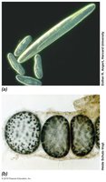
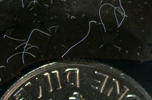
Prokaryotic cell sizes range from 0.2 µm to more than 700 µm in diameter, with average rods measuring 1 x 2 µm. Most eukaryotes are larger, typically 8 µm or more. Some prokaryotes, such as Epulopiscium fishelsoni and Thiomargarita namibiensis, are exceptionally large.
Epulopiscium fishelsoni: Symbiotic in surgeonfish gut, up to 600 µm long.
Thiomargarita namibiensis: Largest known prokaryote, 400–750 µm in diameter.
Surface Area to Volume Ratio
Small cells have a higher surface area-to-volume ratio, which supports greater nutrient exchange and faster growth. This ratio is mathematically described as:
Surface area:
Volume:
Ratio: Surface area/Volume decreases as cell size increases.
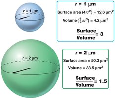
High surface-to-volume ratio allows for rapid evolution due to increased cell division and mutation rates.
Lower Limits of Cell Size
Cells must be large enough to house essential components such as proteins, nucleic acids, and ribosomes. The minimum viable diameter for free-living cells is about 0.2–0.4 µm, commonly observed in open ocean environments and some pathogenic bacteria.
Cytoplasmic Membrane
Structure and Function
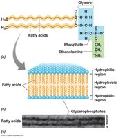
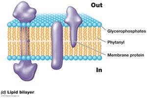
The cytoplasmic membrane is a thin, semi-fluid phospholipid bilayer with embedded proteins. It surrounds the cytoplasm, separates it from the environment, and regulates the traffic of substances via selective permeability. Disruption of the membrane results in cell death.
Phospholipids: Composed of hydrophobic fatty acids and hydrophilic glycerol-phosphate.
Bilayer formation: Fatty acids point inward, hydrophilic portions face the environment or cytoplasm.
Membrane Proteins
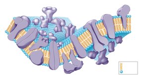
Membrane proteins are classified as integral, transmembrane, lipid-anchored, or peripheral. Integral proteins are permanently embedded, while peripheral proteins associate with membrane surfaces without insertion.
Membrane Structure in Archaea
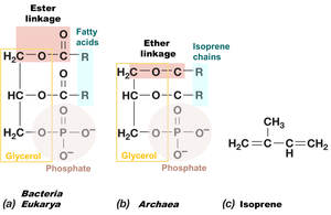
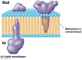
Archaeal membranes differ from bacterial and eukaryotic membranes. Hydrocarbons are derived from isoprene units and attached to glycerol by ether linkages, rather than ester linkages.
Glycerol diethers: Two phytanyl groups attached to glycerol.
Diglycerol tetraether: Two biphytanyl groups attached at both ends, forming a lipid monolayer.
Cyclic rings: Increase membrane rigidity.
Permeability
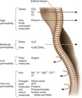
The hydrophobic portion of the membrane acts as a tight barrier to diffusion. Water, though polar, is small enough to pass between phospholipids and is also transported by aquaporins.
Transport Mechanisms in Prokaryotes
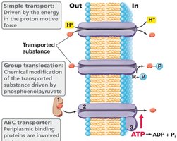
Prokaryotes utilize several transport mechanisms:
Simple transport: Driven by the proton motive force.
Group translocation: Substance is chemically modified during transport.
ABC transporter: Involves substrate-binding protein, membrane transporter, and ATP hydrolyzing protein.
Cell Wall Structure
Gram-Positive vs. Gram-Negative Bacteria
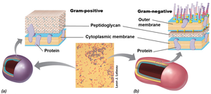
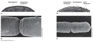
The cell wall protects against lysis and confers shape and rigidity. Gram-positive bacteria have a thick peptidoglycan layer, while gram-negative bacteria have a thin peptidoglycan layer and an outer membrane.
Peptidoglycan Structure
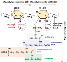
Peptidoglycan is a polymer of glycan tetrapeptide, consisting of N-acetylglucosamine and N-acetylmuramic acid, linked by β-1,4-glycosidic bonds. Peptide bonds connect glycan chains, providing strength in multiple directions.
Lysozyme: Cleaves bonds between sugars, weakening the cell wall.
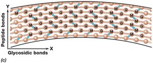
Peptidoglycan Cross-Linking
Gram-negative bacteria form peptide bonds between DAP and D-alanine, while gram-positive bacteria use peptide interbridges, such as five glycine residues in Staphylococcus aureus. Over 100 distinct peptidoglycans have been described.
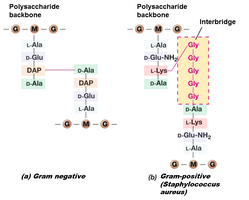
Gram-Positive Cell Wall
Gram-positive cell walls are up to 90% peptidoglycan and contain teichoic acids, which are negatively charged polymers of glycerol or ribitol joined by phosphate groups. These acids are covalently connected to peptidoglycan or plasma membrane lipids.
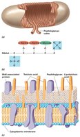
LPS: The Outer Membrane in Gram-Negative Bacteria
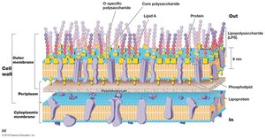
The outer membrane contains phospholipids, proteins, and lipopolysaccharides (LPS). LPS replaces much of the phospholipid in the outer half of the membrane. Lipoproteins connect the peptidoglycan layer to the outer membrane.
Structure of LPS
LPS consists of three main components:
Lipid A: Disaccharide of glucosamine phosphate linked to fatty acids.
Core polysaccharide: Minor variations within genera.
O-specific polysaccharide: Highly variable between species.
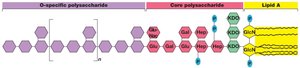
Periplasm & Porins
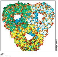
The periplasm is the region between the two membranes, rich in proteins such as hydrolytic enzymes, binding proteins, and chemoreceptors. Porins are proteins that function as channels for solute transport, and may be specific or nonspecific.
Gram Stain Mechanism
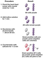
The Gram stain differentiates bacteria based on cell wall structure. Crystal violet forms an insoluble complex with iodine inside the cell. Alcohol dehydrates the thick cell wall of gram-positive cells, trapping the dye, while in gram-negative cells, alcohol extracts the dye complex.
Life Without a Wall
Some prokaryotes naturally lack cell walls, such as Thermoplasma (Archaea) and Mycoplasma (Bacteria). These organisms have tough membranes or live in environments with strict osmotic regulation.
Cell Walls of Archaea
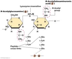
Archaeal cell walls lack peptidoglycan and typically lack an outer membrane. Some have polysaccharide walls or pseudomurein, which is similar to peptidoglycan but contains N-acetyltalosaminuronic acid and β-1,3 linkages, making it lysozyme-insensitive.
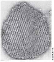
S-Layers in Archaea
S-layers are the most common type of cell wall in Archaea, formed from interlocking layers of protein or glycoprotein. They protect from osmotic lysis, act as a selective sieve, and retain proteins near the surface.
Cell Inclusions
Storage Granules
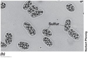
Cell inclusions are granules, crystals, or globules that store substances such as carbon (poly-β-hydroxybutyric acid), inorganic phosphate (polyphosphate chains), and sulfur (in sulfur bacteria).
Gas Vesicles
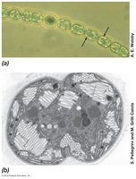
Gas vesicles are spindle-shaped, gas-filled structures that increase buoyancy, allowing cells to adjust their vertical position in water columns.
Capsules & Slime Layers
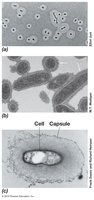
Capsules and slime layers are sticky or slimy materials on the cell surface, consisting of secreted polysaccharide or protein. Capsules adhere firmly, while slime layers are loosely attached. They function in attachment, immune invasion, and protection from dehydration.
Fimbriae and Pili
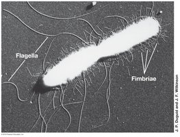
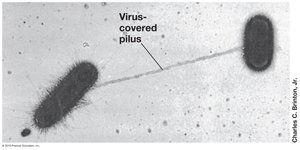
Fimbriae are filamentous proteins that enable cells to stick to surfaces. Pili are longer and thicker, facilitating genetic exchange, adhesion, and twitching motility.
Flagella and Motility
Flagella Arrangements
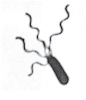
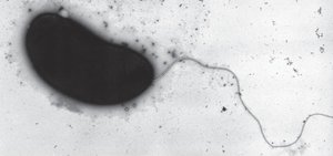
Flagella are threadlike appendages for locomotion. Arrangements include polar (at one or both ends), lophotrichous (tuft at one end), amphitrichous (tufts at both ends), and peritrichous (many locations).
Flagella Structure and Function
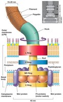
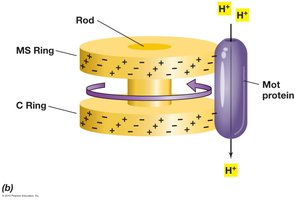
Flagella consist of a basal body (motor), hook, and filament. The basal body anchors the flagella and rotates it, powered by proton movement through Mot proteins.
Archaeal Flagella
Archaeal flagella are thinner, composed of different proteins, and powered by ATP rather than proton motive force. Flagellar motility evolved separately in Bacteria and Archaea.
Swimming Motility
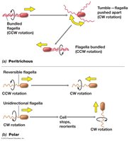
Flagella increase or decrease rotational speed based on proton motive force. Peritrichous flagella move slowly in straight lines, while polar flagella move rapidly and spin.
Gliding Motility
Gliding motility is slower and smoother than flagellar swimming, requiring contact with a solid surface. Mechanisms include secretion of polysaccharide slime, type IV pili twitching, and gliding-specific proteins.
Chemotaxis and Other Taxes
Chemotaxis
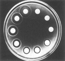
Chemotaxis is movement toward chemical attractants or away from repellents. Chemoreceptors transmit signals internally, altering flagella movement.
Chemotaxis in E. coli
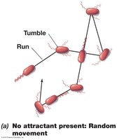
In the absence of attractants, cells move randomly by runs and tumbles. In a gradient, runs toward attractant are longer, and tumbles are less frequent.
Other Taxes
Other directed movements include phototaxis (light), aerotaxis (oxygen), osmotaxis (ionic strength), and hydrotaxis (water).
Bacterial Endospore
Structure and Function
Bacterial endospores are highly differentiated, thick-walled structures produced by certain gram-positive bacteria. They are resistant to heat, chemicals, radiation, and desiccation, and can remain dormant indefinitely.
Endospore Formation and Structure
Endospore formation involves a life cycle from vegetative cell to endospore and back. The mature endospore is located in a characteristic position within the mother cell. The structure includes an exosporium, spore coats, cortex, and core.
Exosporium: Thin protein covering.
Spore coats: Layers of spore-specific proteins.
Cortex: Peptidoglycan, less cross-linked.
Core: Cytoplasmic membrane, cytoplasm, nucleoid, ribosomes, surrounded by a core wall.
Resistance Mechanisms
Endospores are resistant due to protective coats, high levels of dipicolinic acid and calcium, dehydration of the core, and small acid-soluble spore proteins (SASPs) that bind DNA and protect it.
Reactivation
Reactivation occurs in three stages: activation (prepares spore), germination (nutrient detection, water uptake, cortex breakdown), and outgrowth (exit spore coat, return to vegetative state).
Additional info: These notes expand on the original content with academic context, definitions, and examples to ensure completeness and clarity for exam preparation.
