 뒤로
Microbial Structure: Prokaryotic and Eukaryotic Cells
스터디 가이드 - 스마트 노트
 자료에 맞춘 맞춤형 노트, 핵심 정의, 예시, 맥락을 확장해 제공합니다.
Microbial Structure
Three-Domain System
The Three-Domain System, proposed by Carl Woese in 1978, classifies all living organisms into three domains based on differences in ribosomal RNA (rRNA) genes: Bacteria, Archaea, and Eukarya. This system highlights fundamental evolutionary relationships and cellular distinctions.
Bacteria: Includes pathogenic and non-pathogenic prokaryotes.
Archaea: Prokaryotes that live in extreme environments and perform unusual metabolic processes (e.g., extreme halophiles, hyperthermophiles).
Eukarya: Includes animals, plants, fungi, and protists.
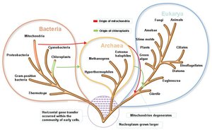
Principal Differences Between Prokaryotic and Eukaryotic Cells
Prokaryotic and eukaryotic cells differ in their structural and functional characteristics. These differences are essential for understanding microbial classification and physiology.
Prokaryotes: One circular chromosome (not membrane-bound), no histones, no membrane-bound organelles, peptidoglycan cell walls (in bacteria), divide by binary fission.
Eukaryotes: Paired chromosomes (membrane-bound), histones present, membrane-bound organelles, polysaccharide cell walls, divide by mitosis using a mitotic spindle.
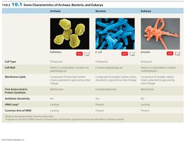
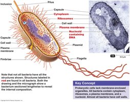
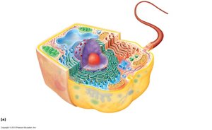
Cell Division: Binary Fission vs. Mitotic Spindle
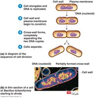
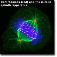
Prokaryotes reproduce by binary fission, a simple process involving cell elongation, DNA replication, and division. Eukaryotes divide by mitosis, which involves a complex mitotic spindle apparatus.
Binary Fission: Cell elongates, DNA replicates, cell wall and membrane constrict, and cells separate.
Mitotic Spindle: Chromosomes are separated by spindle fibers during mitosis.
Summary Table: Principal Differences
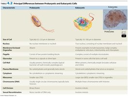
The following table summarizes the key differences between prokaryotic and eukaryotic cells:
Characteristic | Prokaryote | Eukaryote |
|---|---|---|
Size of Cell | 0.2–2.0 µm in diameter | 10–100 µm in diameter |
Nucleus | No nuclear membrane | Nuclear membrane present |
Organelles | Absent | Present |
Cell Wall | Peptidoglycan (bacteria) | Polysaccharides (plants, fungi) |
Division | Binary fission | Mitosis |
Bacterial Cell Structure
What are Bacteria?
Bacteria are prokaryotic microorganisms with over 30,000 described species, and millions more likely undiscovered. They inhabit nearly all environments, reproduce rapidly, and play essential roles in ecosystems and human health.
Majority are harmless; some are pathogenic.
Bacteria can be infected by viruses (bacteriophages).
Important in bioremediation (e.g., oil spill cleanup).
Human skin hosts large numbers of bacteria.
Bacterial Cell Structure
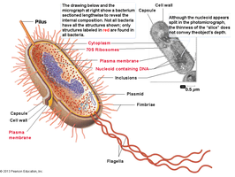
Bacterial cells are typically 0.2–1.0 µm × 2–8 µm in size and exhibit various shapes and arrangements. Their structure includes a cell wall, plasma membrane, cytoplasm, nucleoid, ribosomes, and sometimes external features like flagella, pili, and capsules.
Bacterial Shapes and Arrangements
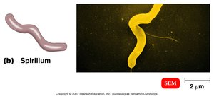
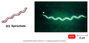
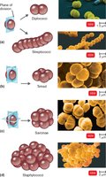
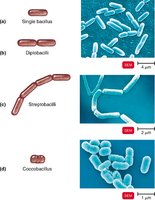
Bacteria are classified by their shapes and cellular arrangements, which are determined by heredity. Most bacteria are monomorphic, but some are pleomorphic.
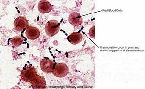
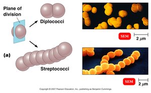
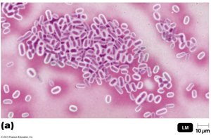
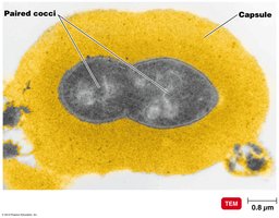
Coccus (Cocci): Spherical shape; arrangements include diplococci (pairs), streptococci (chains), staphylococci (clusters).
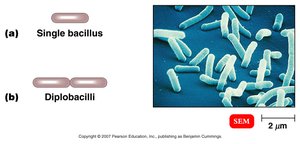
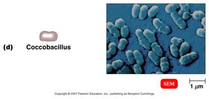
Bacillus (Bacilli): Rod-shaped; arrangements include single bacillus, diplobacilli (pairs), streptobacilli (chains), coccobacillus (short rods).
Spirillum: Helical, rigid structure.
Spirochete: Helical, flexible structure.
Vibrio: Curved rod shape.
Other shapes: Star-shaped, rectangular.
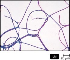
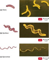
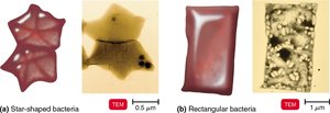
Prokaryotic External Structures
Glycocalyx
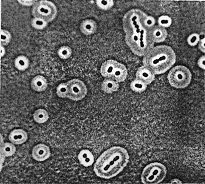
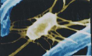
The glycocalyx is an extracellular polysaccharide layer external to the cell wall, aiding in attachment to surfaces and protection from phagocytosis. It exists in two forms:
Capsule: Neatly organized, tightly packed, prevents phagocytosis.
Slime layer: Unorganized, loose.
Flagella
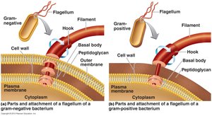
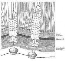
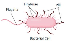
Flagella are long, whip-like appendages used for motility. They are composed of flagellin, attached to a protein hook, and anchored by a basal body. Bacteria can alter the speed and direction of flagellar rotation for movement.
Arrangements: Peritrichous (all over), monotrichous (single, polar), lophotrichous (tuft, polar), amphitrichous (both ends).
Movement: Runs and tumbles, chemotaxis (movement toward/away from chemicals).
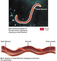
Axial Filament of Spirochetes
Spirochetes possess axial filaments, which are flagella embedded within the membrane, enabling a corkscrew-like movement.

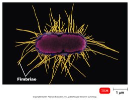
Fimbriae and Pili
Fimbriae are short, hair-like structures that allow bacteria to attach to surfaces. Pili are longer and are used for the transfer of DNA between cells (conjugation).
Bacterial Cell Wall
Structure and Function
The bacterial cell wall provides shape and prevents osmotic lysis. It is composed of peptidoglycan in Gram-positive bacteria, and peptidoglycan plus an outer membrane in Gram-negative bacteria.
Peptidoglycan: Repeating NAG-NAM units cross-linked by polypeptides.
Gram-positive: Contains teichoic acid and lipoteichoic acid.
Gram-negative: Contains an outer membrane with lipopolysaccharide (Lipid A is an endotoxin), O-polysaccharide antigen, and porins.
Atypical Cell Walls
Some bacteria have atypical cell walls:
Mycoplasmas: Lack cell walls, are pleomorphic, contain sterols in plasma membrane.
Acid-fast cell walls: Similar to Gram-positive, but contain waxy lipid (mycolic acid); found in Mycobacterium and Nocardia.
Plasma Membrane and Internal Structures
Plasma Membrane
The plasma membrane is a phospholipid bilayer with embedded proteins and carbohydrates, providing selective permeability and structural integrity.
Contains peripheral, integral, and transmembrane proteins.
Functions in transport, energy generation, and cell signaling.
Internal Structures
Bacterial cytoplasm is the site of biochemical activities and contains the chromosome, ribosomes, enzymes, mRNA, and various inclusions.
Metachromatic granules: Phosphate reserves.
Polysaccharide granules: Energy reserves.
Lipid inclusions: Energy reserves.
Sulfur granules: Energy reserves.
Carboxysomes: CO2 fixation.
Gas vacuoles: Buoyancy.
Magnetosomes: Iron oxide, destroys H2O2.
Endospores
Endospores are highly durable, dehydrated dormant cells that allow bacteria to survive adverse conditions. Sporulation is the formation of spores, and germination is the return to vegetative growth.
Medically significant genera: Bacillus (e.g., anthrax), Clostridium (e.g., tetanus, botulism, gas gangrene, C. difficile).
Eukaryotic Microbes
Structural Complexity
Eukaryotic microbes are structurally more complex and larger than bacteria, with diameters ranging from 10–100 µm. They possess membrane-bound organelles and a nucleus.
Outer Structures
Eukaryotic cell walls are chemically simpler than bacterial cell walls and may contain carbohydrates such as cellulose (plants, algae) or chitin (fungi). Animal cells have a glycocalyx composed of carbohydrates bonded to proteins and lipids.
Flagellum and Cilia
Eukaryotic flagella and cilia are composed of microtubules arranged in a 9+2 array and are made of the protein tubulin. These structures provide motility and are found in various protists and animal cells.
Plasma Membrane and Internal Structures
The eukaryotic plasma membrane is similar to that of bacteria, consisting of a lipid bilayer with proteins and carbohydrates. Eukaryotic cells contain a membrane-bound nucleus, multiple chromosomes associated with histones, and various organelles such as endoplasmic reticulum, Golgi apparatus, lysosomes, mitochondria, and chloroplasts. Cell division occurs by mitosis.
