 뒤로
Microscopy and Microbial Structure: Foundations and Applications in Microbiology
스터디 가이드 - 스마트 노트
 자료에 맞춘 맞춤형 노트, 핵심 정의, 예시, 맥락을 확장해 제공합니다.
Microscopy in Microbiology
Introduction to Microscopy
Microscopy is a fundamental tool in microbiology, enabling the visualization and investigation of microbial structures that are otherwise invisible to the naked eye. The development of various types of microscopes has shaped the history and progress of microbiology, allowing scientists to study cell morphology, structure, and function.
Magnification: The ability of a microscope to enlarge the image of an object beyond its actual size.
Resolution: The minimum distance between two distinct points that can still be distinguished as separate entities.
Applications: Microscopy is used to identify microbial structures, diagnose infections, and investigate cellular processes.
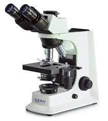
Historical Development of Microscopy
The evolution of microscopy has been marked by key inventions and improvements, each contributing to the ability to observe microorganisms with greater clarity and detail.
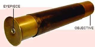
Hans and Zacharias Janssen (1595): Early compound microscope.
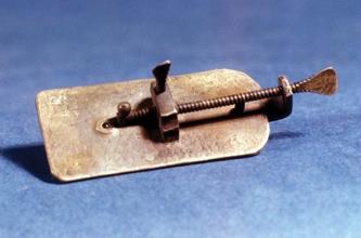
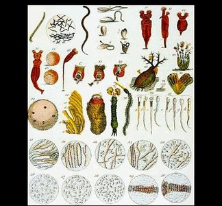
Antonie van Leeuwenhoek (~1673): Single-lens microscope, first to observe bacteria and archaea.
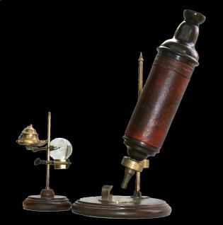
Robert Hooke (~1670): Compound microscope, observed cells in cork.
Modern advancements: Differential interference contrast, phase contrast, super-resolution microscopy.
Key Figures in Microscopy
Antonie van Leeuwenhoek is renowned for his pioneering work in microscopy, being the first to observe and describe microorganisms.
Leeuwenhoek's microscopes: Achieved magnifications of 200-300X, resolving details as small as 1.35 µm.
Impact: Enabled the discovery of bacteria and archaea, laying the foundation for microbiology.

Cell Morphology and Structure
Basic Cell Morphologies
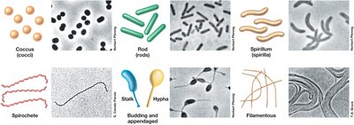
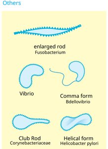
Microbial cells exhibit a wide diversity of shapes and structures, which are important for their identification, classification, and understanding of their functions.
Coccus: Spherical cells.
Rod (Bacillus): Cylindrical cells.
Spirillum: Spiral-shaped cells.
Spirochete: Flexible, spiral-shaped cells.
Stalk, Hypha, Filamentous: Specialized structures for attachment or growth.
Impact of Cell Division on Morphology
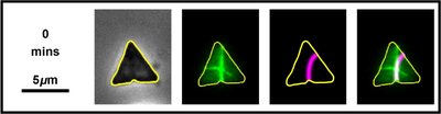
Cell division patterns influence the arrangement and morphology of microbial cells, leading to unique structural forms.
Examples: Chains, clusters, tetrads, and other arrangements resulting from division planes.
Challenges and Techniques in Microbial Visualization
Challenges in Light Microscopy
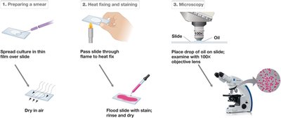
Microbes are often difficult to visualize with standard light microscopy due to their small size and lack of inherent contrast. Various techniques are employed to enhance visibility and distinguish cellular structures.
Simple Dyes/Stains: Basic stains like methylene blue or safranin increase contrast.
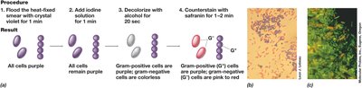
Differential Stains: Gram stain differentiates between Gram-positive and Gram-negative bacteria.
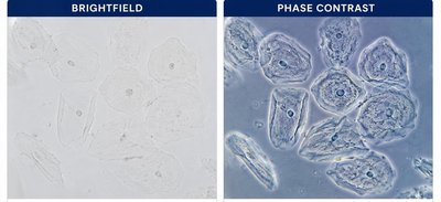
Phase Contrast: Enhances contrast in transparent specimens.
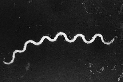
Dark Field Microscopy: Highlights edges and shapes against a dark background.
Fluorescence Microscopy: Uses fluorescent dyes to label specific structures.
Contrast Enhancement Methods
Contrast enhancement is essential for observing live, unstained, or transparent specimens. Each method offers unique advantages for studying microbial structure and function.
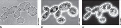
Dark Field Microscopy: Best for observing motility and thin bacteria (e.g., spirochetes).
Phase Contrast Microscopy: Reveals internal structures without staining.
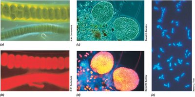
Fluorescence Microscopy: Allows visualization of specific biomolecules and structures.
Benefits of Fluorescence Microscopy
Fluorescence microscopy provides high specificity and clarity, enabling the study of multiple cellular components simultaneously.
Isolate particular wavelengths: Reduces background noise and allows multiplex staining.
Specificity: Targets DNA, lipids, proteins, etc.
Clearer images: Removes signal from non-specific light diffraction.
Advanced Imaging Techniques
Differential Interference Contrast (DIC) and Confocal Microscopy
Advanced light microscopy techniques such as DIC and confocal microscopy provide enhanced resolution and three-dimensional imaging capabilities.
DIC Microscopy: Produces high-contrast images of transparent specimens.
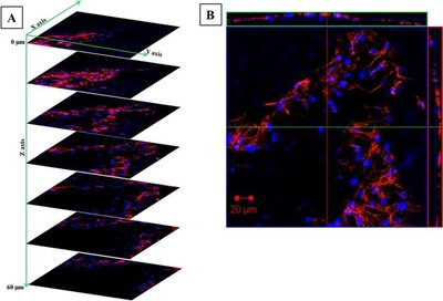
Confocal Microscopy: Uses a pinhole to block out-of-focus light, enabling 3D reconstructions through Z-stacking.
Electron Microscopy
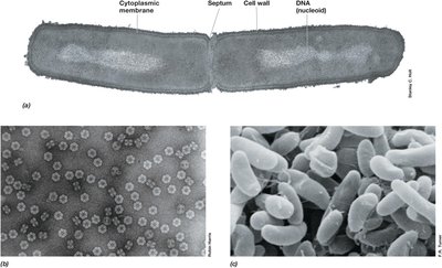
Electron microscopy offers much higher resolution than traditional light microscopy, allowing visualization of ultrastructural details of microbial cells.
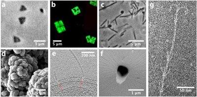
Transmission Electron Microscopy (TEM): Provides detailed internal structures by passing electrons through thin sections.
Scanning Electron Microscopy (SEM): Visualizes surface structures by scanning the specimen with electrons.
Negative Stain: Enhances contrast for TEM imaging.
Cryogenic Electron Tomography (Cryo-ET)
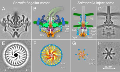
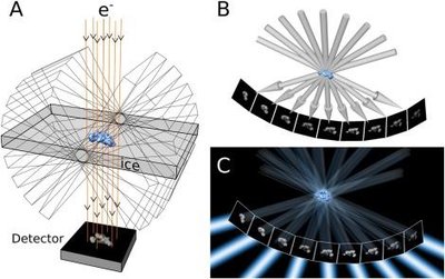
Cryo-ET is a specialized electron microscopy technique that enables three-dimensional reconstructions of cellular and protein structures by imaging samples at multiple tilt angles.
Flash freezing: Preserves native structure without dehydration or fixatives.
3D rendering: Multiple images from different angles are combined to reconstruct the sample.
Applications: Studying cell structures and protein complexes in their native state.
Microbial Structure and Function
Unique Structures of Microbes
Microbes possess specialized structures that confer unique capabilities, such as motility, resistance, and survival under extreme conditions. These structures are often targets for antibiotics, immune responses, or phage infection.
Flagella: Provide motility.
Endospores: Enable survival in harsh environments.
Cell wall: Structural integrity and antibiotic target.
Capsule: Protection from immune system.
Microbe of the Day: Deinococcus radiodurans
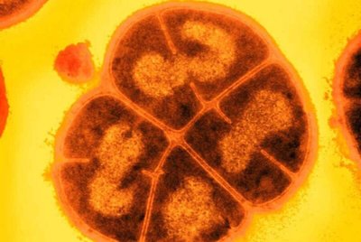
Deinococcus radiodurans is an extremophile bacterium known for its extraordinary resistance to ionizing radiation, dehydration, and other environmental stresses.
Radiation resistance: Survives acute doses up to 5,000 grays (Gy).
Mechanisms: Multiple genome copies, efficient DNA repair, protective proteins.
Applications: Bioremediation, nanotechnology, biomedical research.
Comparing Microbial Eukaryotes, Bacteria, and Archaea
Cellular Properties and Differences
Microbial eukaryotes, bacteria, and archaea exhibit distinct cellular properties, which are important for classification and understanding their biology.
Bacteria: Prokaryotic, peptidoglycan cell wall, diverse morphologies.
Archaea: Prokaryotic, unique cell wall composition, extremophiles.
Eukaryotes: Membrane-bound organelles, complex cell structure.
Feature | Bacteria | Archaea | Eukaryotes |
|---|---|---|---|
Cell Wall | Peptidoglycan | Varied (no peptidoglycan) | Cellulose/chitin (if present) |
Organelles | None | None | Present |
Genome | Circular DNA | Circular DNA | Linear DNA |
Extremophiles | Some | Many | Few |
Summary
Microscopy is essential for the study of microbial structure and function, with a range of techniques available to enhance visualization and resolution. Understanding the unique structures of microbes and their cellular properties is fundamental to microbiology, with implications for antibiotic development, immunity, and biotechnological applications.
