 뒤로
Microbiology Lab 1 Study Guide
스터디 가이드 - 스마트 노트
 자료에 맞춘 맞춤형 노트, 핵심 정의, 예시, 맥락을 확장해 제공합니다.
Microbiology Laboratory Safety and Procedures
General Lab Safety
Proper laboratory safety is essential for preventing contamination and ensuring accurate results in microbiology labs. Adhering to safety protocols protects both the student and the integrity of the experiment.
Personal Protective Equipment (PPE): Always wear a lab coat, gloves, and goggles during laboratory work.
Hand Hygiene: Wash hands before and after lab activities to minimize contamination.
Disinfection: Clean benchtops with disinfectant (e.g., Lysol) before and after experiments.
Proper Storage: Store lab coats and goggles in designated Ziplock bags and cabinets after use.
Prohibited Behaviors: Do not chew gum, bite fingernails, or rest your head on the lab bench.
Waste Disposal: Dispose of used gloves in the appropriate biohazard container.
Lab Attendance and Participation
Active participation and punctuality are mandatory. Attendance is graded, and points are deducted for tardiness, cell phone use, or unexcused absences.
Microscopy and Measurement of Microorganisms
Introduction to Microscopy
Microscopy is a fundamental technique in microbiology for observing the morphology, arrangement, and size of microorganisms. The compound light microscope is commonly used for this purpose.
Oil Immersion Objective (1000x): Provides the highest magnification and resolution for observing bacteria and yeast.
Ocular Micrometer: A scale in the eyepiece used to measure microorganisms. At 1000x, each division typically represents 1 μm.
Microscope Operation and Slide Preparation
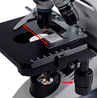
Focusing: Start with a dense area to focus, then move to a region showing the true arrangement of cells.
Artifacts: Slide preparation may cause cells to clump or break apart; always identify the predominant arrangement.
Cleaning: Remove oil from objectives and slides after use with appropriate materials (lens paper, Kimwipe).
Shapes, Arrangements, and Sizes of Microorganisms
Bacterial Morphology
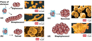
Bacteria are classified by their shapes and arrangements, which are determined by their patterns of cell division and whether daughter cells remain attached.
Cocci (Spherical Bacteria)
Coccus: Spherical or oval-shaped cell (plural: cocci).
Arrangements:
Diplococci: Pairs of cocci.
Streptococci: Chains of cocci.
Tetrads: Groups of four cocci in a square.
Sarcinae: Cubical packets of eight or more cocci.
Staphylococci: Irregular clusters resembling grape bunches.
Bacilli (Rod-Shaped Bacteria)
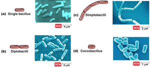
Bacillus: Rod-shaped cell (plural: bacilli).
Arrangements:
Single bacillus: Individual rods.
Diplobacilli: Pairs of rods.
Streptobacilli: Chains of rods.
Coccobacilli: Short, oval rods resembling cocci.
Bacilli divide in one plane only; thus, tetrads, sarcinae, or staphylobacilli do not occur.
Spiral Bacteria
Vibrios: Curved rods.
Spirilla: Thick, rigid spirals.
Spirochetes: Thin, flexible spirals.
Spiral bacteria divide in one plane and do not form clusters or chains.
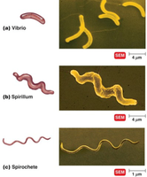
Examples of Bacterial Shapes and Arrangements
Diplococci: Neisseria gonorrhoeae
Streptococci: Streptococcus pyogenes
Tetrads: Micrococcus luteus
Staphylococci: Staphylococcus aureus
Individual bacilli: Escherichia coli
Streptobacilli: Bacillus megaterium
Vibrios: Vibrio species
Spirochetes: Borrelia burgdorferi
Spirilla: Spirillum species
Scientific Names vs. Descriptive Terms
Genus names are italicized and capitalized (e.g., Staphylococcus), while descriptive terms for shape or arrangement are not (e.g., staphylococci).
Yeasts: Eukaryotic Microorganisms
Characteristics of Yeasts
Yeasts are unicellular fungi and are eukaryotic, distinguishing them from prokaryotic bacteria. They are typically spherical, with diameters of 3–5 μm, and reproduce asexually by budding.
Example: Saccharomyces cerevisiae (baker's yeast)
Budding: New cells form as outgrowths from the parent cell.
Measurement of Microorganisms
Using the Ocular Micrometer
The ocular micrometer is a scale in the microscope eyepiece used to measure microorganisms. Calibration is essential for accurate measurement, and at 1000x oil immersion, each division is approximately 1 μm.
Procedure: Focus on the specimen, align the scale, and count divisions to estimate size.
Units: Always report measurements in micrometers (μm).
Comparative Measurement
Measure and compare the size of bacteria, yeast, and a human hair using the ocular micrometer.
Typical sizes:
Bacteria: 0.5–5 μm
Yeast: 3–5 μm
Human hair: 50–100 μm
Study Guide and Skills Assessment
Key Skills for Lab Quiz 1
Identify and describe the different shapes and arrangements of bacteria.
Explain the biological basis for different bacterial arrangements (planes of division, attachment after division).
Recognize artifacts caused by slide preparation.
Measure the size of bacterial and yeast cells using the ocular micrometer, reporting in μm.
Prepare and observe wet mounts of yeast and hair for comparative measurement.
Sample Table: Bacterial Shapes and Arrangements
Shape | Arrangement | Example Organism |
|---|---|---|
Coccus | Diplococci | Neisseria gonorrhoeae |
Coccus | Streptococci | Streptococcus pyogenes |
Coccus | Tetrads | Micrococcus luteus |
Coccus | Staphylococci | Staphylococcus aureus |
Bacillus | Individual | Escherichia coli |
Bacillus | Streptobacilli | Bacillus megaterium |
Spiral | Vibrio | Vibrio species |
Spiral | Spirochete | Borrelia burgdorferi |
Spiral | Spirillum | Spirillum species |
Additional Info
Artifacts in bacterial arrangements are often due to mechanical disruption during slide preparation.
Proper identification of bacterial morphology is crucial for clinical diagnosis and taxonomy.
