 뒤로
Chapter 20: Management of Patients With Nonacute Disorders of the Chest and Lower Respiratory Tract and Disorders of the Pulmonary Vasculature
스터디 가이드 - 스마트 노트
 자료에 맞춘 맞춤형 노트, 핵심 정의, 예시, 맥락을 확장해 제공합니다.
Chapter 20: Management of Patients With Nonacute Disorders of the Chest and Lower Respiratory Tract and Disorders of the Pulmonary Vasculature
Pulmonary Tuberculosis
Tuberculosis (TB) is a chronic infectious disease primarily affecting the lung parenchyma, but it can also spread to other organs such as the meninges, kidneys, bones, and lymph nodes. It is caused by Mycobacterium tuberculosis, an acid-fast aerobic rod sensitive to heat and ultraviolet light. TB remains a global public health issue, closely associated with poverty, malnutrition, overcrowding, and inadequate health care.
Transmission: TB spreads via airborne droplet nuclei released during talking, coughing, or sneezing.
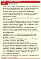
Risk Factors: Close contact with active TB cases, immunocompromised status, substance use disorder, inadequate health care, preexisting medical conditions, immigration from high-prevalence countries, living in group settings, and health care workers performing high-risk activities.
Pathophysiology: Inhaled mycobacteria reach the alveoli, multiply, and spread via lymphatic and blood systems. The immune response forms granulomas, which may become fibrous and calcified. Active disease can develop due to compromised immunity or reinfection.
Clinical Manifestations: Symptoms are insidious and chronic, including low-grade fever, cough, night sweats, fatigue, weight loss, and hemoptysis. Extrapulmonary disease is more common in patients with HIV.
Assessment and Diagnostic Findings
Diagnosis of TB involves a combination of clinical, radiological, and laboratory findings.
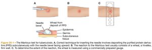
Skin Test: The Mantoux method uses purified protein derivative (PPD) injected intracutaneously. Induration is measured 48–72 hours later. A significant reaction indicates past exposure or vaccination with BCG, but does not confirm active disease.
Blood Tests: Interferon-gamma release assays (IGRAs) such as QuantiFERON-TB Gold Plus and T-SPOT are preferred for patients with BCG vaccine or those unlikely to return for skin test reading.
Sputum Culture: Presence of acid-fast bacilli (AFB) on smear may indicate disease, but culture confirms diagnosis. Initial isolates should be tested for drug resistance.
Imaging: Chest x-ray typically shows lesions in the upper lobes.
Medical Management of Tuberculosis
Treatment of TB requires prolonged administration of anti-TB agents to ensure eradication and prevent relapse. Drug resistance is a major concern, with multidrug-resistant (MDR), pre-extensively drug-resistant (pre-XDR), and extensively drug-resistant (XDR) TB types.
Combination Therapy: Medications such as isoniazid and rifampin are often combined to improve adherence and efficacy.
Duration: Standard treatment lasts 4–9 months, depending on drug susceptibility and patient response.
Monitoring: Initial isolates should be tested for drug resistance.
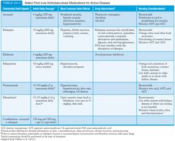
Drug Name | Adult Daily Dosage | Most Common Side Effects | Drug Interaction | Nursing Considerations |
|---|---|---|---|---|
Isoniazid | 5 mg/kg (max 300 mg) | Hepatitis, peripheral neuropathy, lupus-like syndrome | Antacids, alcohol | Monitor liver function, educate on side effects |
Rifampin | 10 mg/kg (max 600 mg) | Hepatitis, orange urine, GI upset | Oral contraceptives, anticoagulants | Monitor liver function, educate on urine discoloration |
Ethambutol | 15–25 mg/kg | Optic neuritis, rash | None significant | Monitor vision, educate on symptoms |
Pyrazinamide | 15–30 mg/kg | Hepatitis, arthralgia | None significant | Monitor liver function, educate on joint pain |
Nursing Management of Tuberculosis
Nursing care focuses on promoting airway clearance, encouraging adherence to treatment, supporting activity and nutrition, and preventing transmission. Education on medication schedule, side effects, and hygiene measures is essential.
Increase fluid intake and use postural drainage.
Plan progressive activity schedule and provide nutritional support.
Educate on hygiene, report to health department, monitor for miliary TB.
Lung Abscess
Pathophysiology and Clinical Manifestations
A lung abscess is a localized collection of pus caused by microbial infection, often as a complication of bacterial pneumonia or aspiration of oral anaerobes. It may also result from tumors, foreign bodies, necrotizing pneumonias, TB, pulmonary embolism, or trauma.
Pathophysiology: Most abscesses are complications of bacterial pneumonia or aspiration. The site is influenced by gravity and patient position. Abscesses become surrounded by fibrous tissue and may extend to bronchus or pleural space.
Clinical Manifestations: Symptoms range from mild productive cough to acute illness, including fever, productive cough with foul-smelling sputum, leukocytosis, pleurisy, dull chest pain, dyspnea, weakness, anorexia, and weight loss.
Assessment and Diagnostic Findings
Diagnosis is confirmed by chest x-ray, sputum culture, and fiberoptic bronchoscopy. Chest x-ray shows infiltrate with air-fluid level; CT scan may provide detailed images.
Physical exam may reveal dullness on percussion, decreased breath sounds, pleural friction rub, and crackles.
Medical and Nursing Management
Treatment is guided by history, physical exam, chest x-ray, and sputum culture. Chest physiotherapy and postural drainage are used for adequate drainage. IV antimicrobial therapy is based on sputum culture results, with standard treatment including ampicillin–sulbactam. Duration is typically 3 weeks or longer.
Monitor for adverse effects, initiate chest physiotherapy, educate on deep-breathing and coughing exercises, encourage high-protein, high-calorie diet, and provide emotional support.
Sarcoidosis
Pathophysiology and Clinical Manifestations
Sarcoidosis is an inflammatory, multisystem, granulomatous disease of unknown etiology, most commonly affecting adults aged 30–50. It is more frequent in Black Americans and those of Scandinavian descent.
Pathophysiology: Thought to be a hypersensitivity response to exogenous agents (bacteria, fungi, virus, chemicals), resulting in formation of noncaseating granulomas.
Clinical Manifestations: Insidious onset with dyspnea, cough, hemoptysis, congestion, uveitis, joint pain, fever, granulomatous lesions, fatigue, anorexia, and weight loss.
Assessment and Diagnostic Findings
Diagnosis is confirmed by biopsy showing noncaseating granulomas. Chest x-rays and CT scans assess pulmonary adenopathy, showing hilar adenopathy and miliary/nodular lesions. Pulmonary function tests may show restriction; arterial blood gas may show hypoxemia and hypercapnia.
Medical and Nursing Management
Many patients achieve remission without treatment. Corticosteroids (prednisone) are used for symptom relief and respiratory impairment control. Immune modulators and monoclonal antibody agents may be added for refractory cases.
Educate on corticosteroid effects and usage, encourage follow-ups, assess disease progression, and refer to support resources.
Pleural Disorders
Pleurisy
Pleurisy is inflammation of both layers of the pleurae (parietal and visceral), often developing with pneumonia, upper respiratory tract infection, TB, or collagen disease. Symptoms include severe, sharp, knifelike pain intensified on inspiration.
Pleuritic pain worsens with deep breath, coughing, or sneezing, usually occurs on one side, and may radiate to shoulder or abdomen.
Pain decreases as pleural fluid develops.
Pleural Effusion
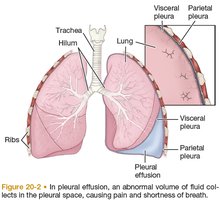
Pleural effusion is the collection of fluid in the pleural space, secondary to other diseases such as heart failure, TB, or pneumonia. Normal pleural fluid acts as a lubricant for pleural surfaces.
Pathophysiology: Fluid accumulation can be clear, bloody, or purulent. Transudate is clear fluid from heart failure; exudate results from inflammation or tumors.
Clinical Manifestations: Symptoms depend on underlying disease and effusion size, including fever, chills, pleuritic chest pain, dyspnea, and cough.
Assessment: Physical exam shows decreased/absent breath sounds, decreased fremitus, and dull percussion. Diagnostic tests include chest x-ray, CT, and thoracentesis.
Medical Management: Treatment aims to discover underlying cause, prevent fluid reaccumulation, and relieve discomfort. Thoracentesis removes fluid and relieves dyspnea; chest tube or chemical pleurodesis may be required for recurrent effusions.
Empyema
Empyema is the accumulation of thick, purulent fluid in the pleural space, often with fibrin development and loculated area. It is usually a complication of bacterial pneumonia or lung abscess.
Pathophysiology: Initial pleural fluid is thin with low leukocyte count, progressing to fibropurulent stage, and finally thick exudative membrane enclosing the lung.
Clinical Manifestations: Symptoms are similar to acute respiratory infection or pneumonia, including fever, night sweats, pleural pain, cough, dyspnea, anorexia, and weight loss.
Assessment: Chest auscultation reveals decreased/absent breath sounds, dullness on percussion, and decreased fremitus. Diagnosis is confirmed by chest CT and diagnostic thoracentesis.
Medical Management: Treatment objectives are to drain the pleural cavity and achieve complete lung expansion. Methods include thoracentesis, tube thoracostomy, open chest drainage, and long-term antibiotics.
Pulmonary Vascular Disorders
Pulmonary Hypertension (PH)
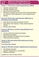
Pulmonary hypertension is elevated pulmonary arterial pressure leading to right ventricular heart failure. It is classified into five groups based on underlying mechanism.
Group 1: Pulmonary arterial hypertension (PAH)
Group 2: PH due to left-sided heart disease
Group 3: PH due to chronic lung diseases/hypoxemia
Group 4: PH associated with pulmonary artery obstructions
Group 5: PH with unclear multifactorial mechanisms
Pathophysiology and Clinical Manifestations
Risk factors include collagen vascular disease, congenital heart disease, chronic stimulant use, portal hypertension, and HIV. Vascular injury leads to endothelial and vascular smooth muscle dysfunction, increasing pulmonary artery pressure and affecting right ventricular function.
Symptoms: Dyspnea (initially with exertion, later at rest), substernal chest pain, weakness, fatigue, syncope, hemoptysis, signs of right-sided heart failure (peripheral edema, ascites, distended neck veins, liver engorgement, crackles, heart murmur).
Assessment and Diagnostic Findings
Diagnosis is confirmed by history, physical exam, chest x-ray, pulmonary function studies, ECG, echocardiogram, and right heart catheterization. Mean pulmonary artery pressure >20 mm Hg and wedge pressure ≤15 mm Hg are diagnostic criteria.
Medical and Pharmacologic Management
Primary goal is to manage the underlying condition. Supportive therapies include diuretics, oxygen, anticoagulation, digoxin, and exercise training. Pharmacologic advances include calcium channel blockers, prostanoids, endothelin receptor antagonists, phosphodiesterase-5 inhibitors, and soluble guanylate cyclase stimulants.
Vasoreactivity test during cardiac catheterization identifies best medication.
Lung transplantation and atrial septostomy are surgical options for refractory cases.
Nursing Management
Identify at-risk patients for early treatment, administer oxygen therapy, educate on home oxygen therapy, prostanoid administration, and provide emotional and psychosocial support.
Pulmonary Heart Disease (Cor Pulmonale)
Cor pulmonale results from pulmonary hypertension, causing right heart enlargement and right-sided heart failure.
Pulmonary Embolism (PE)
PE is the obstruction of the pulmonary artery or branches by thrombus, usually originating in the venous system or right heart. Deep vein thrombosis (DVT) and venous thromboembolism (VTE) are related conditions.
Occupational Lung Disease: Pneumoconioses
Definition and Types
Pneumoconiosis is a general term for lung disease caused by inhaled dusts, resulting in pulmonary fibrosis and parenchymal changes. Types include asbestosis, silicosis, and coal workers pneumoconiosis (black lung disease).
Pathophysiology: Non-neoplastic alteration of the lung from inhalation of mineral/inorganic dust.
Exposure: Extended exposure to irritating/toxic substances; severe single exposures may lead to chronic lung disease.
Prevalence: Number one work-related illness in the United States.
Prevention and Management
Diseases are not curable but preventable. Nurses advocate for employees by promoting measures to reduce worker exposure, including protective devices, screening, and monitoring.
Key assessment aspects: job activities, exposure levels, hygiene, time frame, smoking history, effectiveness of respiratory protection.
Veterans Considerations
Veterans may be exposed to pollutants from burn pits, sand storms, and car bombings, leading to respiratory disorders ranging from asthma to constrictive bronchiolitis. Assessment of exposure history is crucial.
Lung Cancer
Classification and Risk Factors
Lung cancer is most commonly caused by inhaled carcinogens from cigarette smoke. Classifications include small cell lung cancer (SCLC) and non–small cell lung cancer (NSCLC).
Risk Factors: Tobacco smoke, electronic nicotine delivery systems, second-hand smoke, environmental and occupational hazards, genetic mutations.
Clinical Manifestations: Cough or change in chronic cough, dyspnea, hemoptysis.
Assessment: Chest x-ray, CT scan, biopsy.
Medical Management
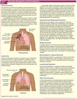
Treatment aims to provide cure if possible, using surgery, radiation, and chemotherapy. Thoracotomy may be required. Pre- and postoperative nursing management is essential. ----------------------------------------
