 뒤로
Observing Microorganisms Through a Microscope: Microscopy and Staining Techniques
스터디 가이드 - 스마트 노트
 자료에 맞춘 맞춤형 노트, 핵심 정의, 예시, 맥락을 확장해 제공합니다.
Microscopes and Magnification
Introduction to Microscopy
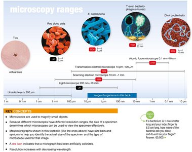
Microscopy is fundamental in microbiology, enabling the observation of microorganisms that are invisible to the naked eye. The range of microscopes and their magnification capabilities allow scientists to study cells, bacteria, viruses, and molecular structures.
Microscopes are instruments used to magnify small objects, making them visible for study.
Resolution refers to the ability to distinguish two points as separate entities; higher resolution means finer detail.
Different microscopes have different resolution ranges, affecting the size of specimens that can be observed.
Types of Microscopes
Light microscopes use visible light to observe specimens and are suitable for cells and bacteria.
Electron microscopes (transmission and scanning) provide higher resolution for viruses and molecular structures.
Atomic force microscopes can visualize structures at the nanometer scale.
Compound Light Microscope
Principal Parts and Functions
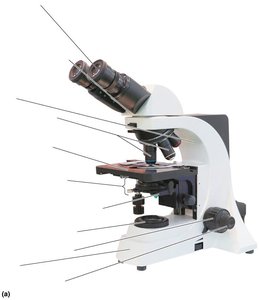
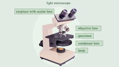
The compound light microscope is the most common instrument in microbiology labs. It uses a series of lenses to magnify specimens.
Ocular lens (eyepiece): Remagnifies the image formed by the objective lens.
Objective lenses: Primary lenses that magnify the specimen.
Stage: Holds the microscope slide in position.
Condenser: Focuses light through the specimen.
Diaphragm: Controls the amount of light entering the condenser.
Illuminator: Light source.
Coarse and fine focusing knobs: Adjust focus.
Magnification and Path of Light
Total magnification is calculated as:
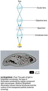
The path of light travels from the illuminator, through the condenser, specimen, objective lens, body tube, and ocular lens to the eye.
Resolution and Refraction
Resolution is the ability to distinguish two points; shorter wavelengths of light provide greater resolution.
Refractive index measures the light-bending ability of a medium. Immersion oil is used to prevent light refraction and loss, improving resolution.
Types of Light Microscopy
Brightfield Microscopy
Brightfield microscopy is the standard method, where dark objects are visible against a bright background.
Light passes through the specimen, and internal structures are visible.
Used for stained specimens.
Darkfield Microscopy
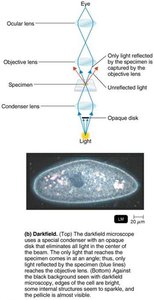
Darkfield microscopy enhances contrast by making light objects visible against a dark background.
An opaque disk in the condenser blocks direct light; only light reflected by the specimen enters the objective lens.
Useful for observing live, unstained specimens.
Phase-Contrast Microscopy
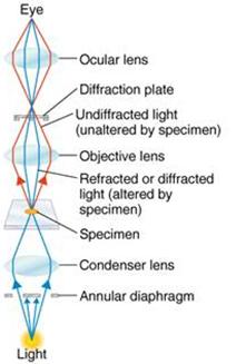
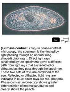
Phase-contrast microscopy allows examination of living organisms and internal cell structures without staining.
Combines direct and diffracted light rays to form an image.
Shows greater differentiation of internal structures.
Preparing Microbes for Staining
Smear Preparation
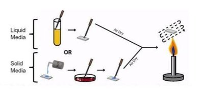
Preparing a smear is the first step in staining bacteria.
A thin film of microorganisms is spread over a slide.
Microorganisms are fixed to the slide by heat, which kills them.
Live and unstained specimens have little contrast; staining is used to emphasize structures.
Staining Techniques
Types of Stains
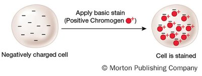
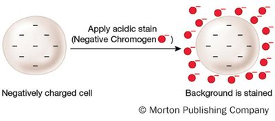
Stains consist of a positive and negative ion, one of which is colored (chromophore).
Basic dye: Chromophore is a cation; stains the cell.
Acidic dye: Chromophore is an anion; stains the background (negative staining).
Simple Stains
Simple stains use a single basic dye to highlight the entire microorganism, making cell shapes and structures visible.
A mordant may be used to intensify the stain or increase its affinity for the specimen.
Mordants can also coat structures (e.g., flagella) to make them easier to see.

Differential Stains
Differential stains react differently with various bacteria, allowing distinction between types. The most common are the Gram stain and Acid-fast stain.
Gram Stain
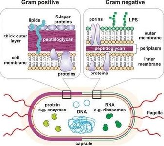
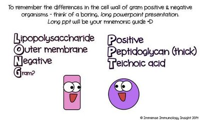
The Gram stain differentiates bacteria based on cell wall structure.
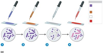
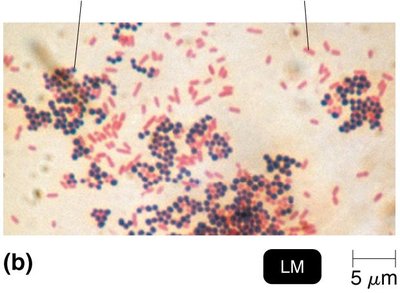
Gram-positive bacteria: Thick peptidoglycan layer; retain crystal violet stain (appear purple).
Gram-negative bacteria: Thin peptidoglycan layer and outer membrane; lose crystal violet and take up safranin (appear pink/red).
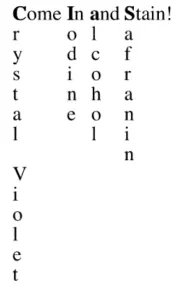
Steps: Application of crystal violet, iodine (mordant), alcohol wash (decolorization), and safranin (counterstain).
Acid-Fast Stain
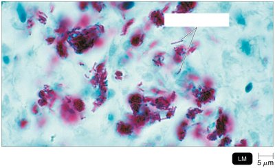
The acid-fast stain is used for bacteria with waxy cell walls (e.g., Mycobacterium, Nocardia).
Primary stain: carbolfuchsin (red).
Decolorizing agent: acid-alcohol.
Counterstain: methylene blue.
Acid-fast bacteria retain red; non–acid-fast bacteria appear blue.
Special Stains
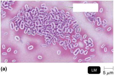
Capsule Stain
Capsule stains are used to visualize gelatinous coverings (capsules) that do not accept most dyes.
Negative staining with India ink or nigrosin contrasts the background, making the capsule appear as a halo.
Endospore Stain
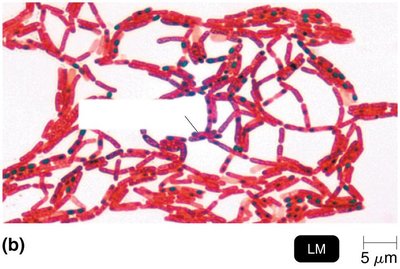
Endospore stains identify resistant, dormant structures within cells.
Primary stain: malachite green (with heat).
Decolorize with water.
Counterstain: safranin.
Endospores appear green within red or pink cells.
Heat/steam acts as the mordant.
Flagella Stain
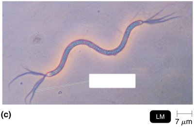
Flagella stains make structures of locomotion visible by thickening them with a mordant and carbolfuchsin.
Flagella are otherwise too thin to be seen with light microscopy.
Summary Table: Staining Methods and Their Applications
Staining Method | Purpose | Key Steps | Result |
|---|---|---|---|
Simple Stain | Visualize cell shape and structure | Single basic dye, optional mordant | Entire cell stained |
Gram Stain | Differentiate Gram-positive and Gram-negative bacteria | Crystal violet, iodine, alcohol, safranin | Purple (positive), Pink (negative) |
Acid-Fast Stain | Identify acid-fast bacteria | Carbolfuchsin, acid-alcohol, methylene blue | Red (acid-fast), Blue (non–acid-fast) |
Capsule Stain | Visualize capsules | Negative staining with India ink/nigrosin | Capsule appears as halo |
Endospore Stain | Identify endospores | Malachite green (heat), water, safranin | Green endospores in red cells |
Flagella Stain | Visualize flagella | Mordant, carbolfuchsin | Flagella visible |
Key Terms and Concepts
Microscope: Instrument for magnifying small objects.
Magnification: Increase in apparent size of an object.
Resolution: Ability to distinguish two points as separate.
Stain: Dye used to color cells or cell structures.
Mordant: Substance that intensifies a stain or binds it to a specimen.
Gram-positive/Gram-negative: Classification based on cell wall structure and staining response.
Acid-fast: Bacteria resistant to decolorization by acid-alcohol.
Capsule, Endospore, Flagella: Specialized structures visualized by special stains.
Additional info: The notes above expand on brief points with academic context, definitions, and examples to ensure completeness and clarity for microbiology students.
