 뒤로
Lab 5: Preparation of Smears and Simple Staining in Microbiology
스터디 가이드 - 스마트 노트
 자료에 맞춘 맞춤형 노트, 핵심 정의, 예시, 맥락을 확장해 제공합니다.
Preparation of Smears and Simple Staining
Introduction to Simple Staining
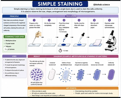
Simple staining is a fundamental microbiological technique used to visualize bacterial cells under a microscope. By applying a single basic dye, the size, shape, arrangement, and morphology of microorganisms can be observed. This technique is essential for differentiating between bacterial species and understanding their structural characteristics.
Chemistry of Staining
Why Staining Works
Staining relies on the interaction between charged dye molecules and the bacterial cell surface. Most bacterial cells possess a net negative charge, which attracts positively charged (cationic) dyes, allowing for effective staining and visualization.
Basic (Cationic) Stains: The chromophore (colored ion) carries a positive charge and binds to the negatively charged bacterial cell surface. Examples include methylene blue, crystal violet, malachite green, safranine, and fuchsin.
Acidic (Anionic) Stains: The chromophore carries a negative charge and is repelled by the bacterial cell surface, making these dyes unsuitable for direct staining of bacteria. Examples include eosin, picric acid, rose Bengal, and acid fuchsin.
Key Concept: The effectiveness of staining is based on ionic interactions between the dye and the cell surface.
Ionic Bonds and Salts in Staining
Ionic bonds are formed when one atom donates electrons to another, resulting in positively and negatively charged ions that attract each other. Stains used in microbiology are typically salts, consisting of a positive and a negative ion, one of which is the chromophore.
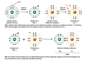
Example: Methylene blue+ (cation) + Cl− (anion)
Preparation of Bacterial Smears
Materials and Cultures
Wash bottle of distilled water
Glass slides
Inoculating loop
95% methanol (optional for chemical fixation)
Methylene blue
Staphylococcus epidermidis (from solid medium)
Bacillus megaterium (from broth culture)
Smear Preparation Steps
From a Slant (Solid Medium): Place a drop of water on the slide, aseptically transfer a small amount of growth, and mix to create a light suspension.
From a Broth (Liquid Medium): Transfer 2–3 loopfuls of broth directly onto the slide (no water needed).
Spread the sample into a thin circle and allow it to air-dry at room temperature.
Heat-Fixing: Pass the slide through a flame 3–5 times to kill bacteria and adhere them to the slide.
Definitions:
Smear: A thin film of material containing microorganisms spread over a slide.
Fixation: The process of killing bacteria and attaching them to the slide, either by heat or chemicals (e.g., methanol).
Staining Procedure
Steps for Simple Staining
Hold the slide with a clothespin, forceps, or slide holder.
Flood the smear with methylene blue for 30–60 seconds.
Rinse gently with distilled water.
Blot dry with bibulous paper (do not rub).
Examine under the microscope: start with low power (10x), then high-dry (40x), and finally oil immersion (100x).
Record observations by sketching each organism at oil immersion.
Results and Interpretation
Expected Observations
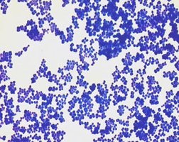
Staphylococcus epidermidis: Gram-positive cocci in grape-like clusters.
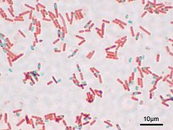
Bacillus megaterium: Large, rod-shaped, spore-forming bacteria, often found singly or in chains.
Bacterial Shapes and Arrangements
Basic Bacterial Shapes
Coccus (cocci): Spherical or round-shaped bacteria.
Bacillus (bacilli): Rod-shaped bacteria.
Spiral: Curved or twisted bacteria.
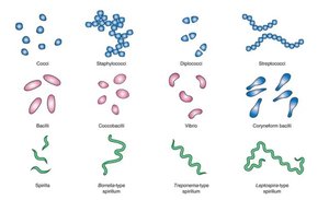
Bacterial Arrangements
Bacilli: Single, diplobacilli (pairs), streptobacilli (chains).
Cocci: Diplococcus (pairs), streptococcus (chains), tetrads (groups of 4), sarcinae (cubes of 8), staphylococcus (clusters).
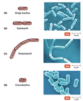
Genus Bacillus and Staphylococcus
Bacillus
Aerobic or facultatively anaerobic, spore-forming rods.
Environmental survivors; some species are important in industry and medicine.
Examples: B. subtilis (probiotics), B. anthracis (anthrax), B. cereus (food poisoning).
Staphylococcus
Gram-positive cocci in grape-like clusters.
Commonly found on skin and nasal flora; salt-tolerant, facultative anaerobes, non-spore-forming.
Important species: S. epidermidis (skin), S. saprophyticus (urogenital), S. aureus (pathogenic).
Review Questions and Applications
Key Questions
Value of a Simple Stain: Allows visualization of cell shape, size, and arrangement.
Purpose of Heat-Fixing: Kills bacteria and adheres them to the slide, preventing washing off during staining.
Methanol Fixation: Alcohol denatures proteins and causes cells to adhere to the slide by dehydration and coagulation.
Excessive Heat-Fixing: Can distort cell morphology and destroy cellular structures.
Conclusions
Bacillus: Bacillus megaterium is the bacillus (rod-shaped) bacterium.
Relative Size: Bacillus megaterium is larger than Staphylococcus epidermidis.
Summary Table: Basic vs. Acidic Dyes
Type of Dye | Charge on Chromophore | Examples | Binding to Bacteria |
|---|---|---|---|
Basic (Cationic) | Positive | Methylene blue, crystal violet, safranine | Binds to negatively charged cell surface |
Acidic (Anionic) | Negative | Eosin, acid fuchsin | Repelled by cell surface; stains background |
Additional info: Simple staining is foundational for more advanced staining techniques, such as Gram staining and differential staining, which further classify bacteria based on cell wall properties.
