뒤로
Viruses and Prions: Structure, Classification, Replication, and Clinical Principles
스터디 가이드 - 스마트 노트
 자료에 맞춘 맞춤형 노트, 핵심 정의, 예시, 맥락을 확장해 제공합니다.
Viruses and Prions: Basic Principles
Viruses as Nonliving Pathogens
Viruses are acellular, submicroscopic infectious agents that require a host cell for replication. They are considered obligate intracellular pathogens and are studied in the field of virology. Over 5,000 mammal-infecting viral species have been described, with many more uncharacterized. Most viruses that infect humans are harbored in other animals.
Size: Extremely small (20–400 nm)
Acellular: Lack cellular structure
Obligate intracellular: Cannot replicate outside a host cell
Comparison of Viruses, Prokaryotes, and Eukaryotes
Viruses differ fundamentally from prokaryotic and eukaryotic cells in structure, replication, and metabolism.
Feature | Viruses | Prokaryotes | Eukaryotes |
|---|---|---|---|
Cellular structure | No | Yes | Yes |
Genetic material | DNA or RNA | DNA | DNA |
Replication | Host-dependent | Binary fission | Mitosis/meiosis |
Metabolism | No | Yes | Yes |
Viral Structure and Genomic Features
Virion and Capsid
A virion is a single, infectious virus particle. It consists of an exterior protective protein capsid and genetic material (DNA or RNA). The capsid is made of capsomere subunits and accounts for most of the virion's mass.
Capsid: Protein shell protecting the genome
Capsomeres: Subunits forming the capsid
Capsid Structures
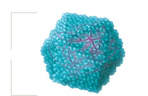
Most animal viruses have either helical or icosahedral capsids. Deviations are classified as complex capsids.
Helical capsid: Hollow tube-like structure
Icosahedral capsid: Three-dimensional polygon
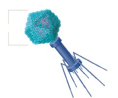
Complex capsid: Found in bacteriophages, often with additional structures for genome injection

Viral Envelopes
Some viruses possess a lipid-based envelope surrounding the capsid, acquired from the host cell membrane during budding. Naked viruses lack this envelope and are released by cell lysis.
Enveloped viruses: Arise from budding
Naked viruses: Arise from cell lysis
Bacteriophages: Always naked
Viral Spikes (Peplomers)
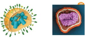
Many viruses have spikes (glycoprotein extensions) protruding from the capsid or envelope, facilitating attachment and entry into host cells. Spikes bind to specific host cell factors.
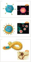
Influenza viruses frequently mutate their spike proteins, leading to antigenic drift and shift. The main spikes are hemagglutinin (HA) and neuraminidase (NA).
Viral Genomes
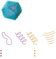
Viral genomes are highly diverse and typically contain fewer than 300 genes. They encode capsomere proteins, replication enzymes, and structural factors. Genomes may be DNA or RNA, single or double-stranded, segmented or non-segmented, circular or linear.
DNA viruses: Often double-stranded, can be circular or linear
RNA viruses: Often single-stranded, can be linear or segmented
Viral Genome Replication Strategies
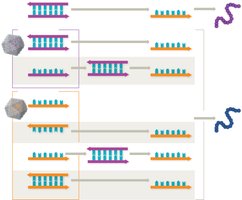
Viruses use various strategies to produce mRNA for protein synthesis, depending on their genome type.
dsDNA viruses: Use host RNA polymerases for transcription
ssDNA viruses: Converted to dsDNA before transcription
ssRNA+ viruses: Genome acts as mRNA, directly translated
ssRNA- viruses: Genome is complementary to mRNA, transcribed by viral RNA-dependent RNA polymerases
Retroviruses: ssRNA genome converted to DNA by reverse transcriptase, integrated into host genome
dsRNA viruses: Transcribed to make mRNA, requires viral RNA-dependent RNA polymerases
Viral Genome Evolution
Mutation and Reassortment
Viruses mutate rapidly due to quick replication and lack of proofreading in RNA polymerases. Mutations can be neutral, beneficial, or deleterious. Reassortment occurs when two viral strains coinfect a host cell, producing new strains.
Antigenic drift: Minor changes in viral antigens (e.g., influenza spikes)
Antigenic shift: Major genetic reassortment, can lead to pandemics
Classification and Naming of Viruses
Classification Criteria
The International Committee on Taxonomy of Viruses (ICTV) classifies viruses based on:
Type of nucleic acid (DNA or RNA)
Capsid symmetry (helical, icosahedral, complex)
Presence or absence of envelope
Genome architecture (ssDNA, dsDNA, ssRNA, etc.)
Medically Important Viral Families
Viruses are grouped into families based on genome type, capsid shape, and envelope status. Examples include:
DNA virus families: Papillomaviridae, Parvoviridae, Adenoviridae, Hepadnaviridae, Poxviridae, Herpesviridae
RNA virus families: Calciviridae, Picornaviridae, Paramyxoviridae, Filoviridae, Rhabdoviridae, Bunyaviridae, Orthomyxoviridae, Arenaviridae, Flaviviridae, Retroviridae, Togaviridae, Reoviridae, Coronaviridae
Host Range and Tropism
Host range is the collection of species a virus can infect. Tropism refers to the specificity for certain tissues or cell types, determined by viral surface factors.
Virus Sizes
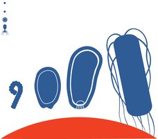
Viruses vary greatly in size, from 30 nm (rhinovirus, poliovirus) to 1,500 nm (pithovirus).
Naming Conventions
Viruses are not assigned to domains or kingdoms. The highest taxon is phylum, followed by order, family, genus, and species.
Viral Replication Pathways
Bacteriophage Replication
Bacteriophages replicate via two main pathways: lytic and lysogenic.
Lytic pathway: Immediate virion production, host cell lysis
Lysogenic pathway: Viral genome integrates into host genome as a prophage, replicated with host DNA, can later enter lytic cycle
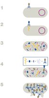
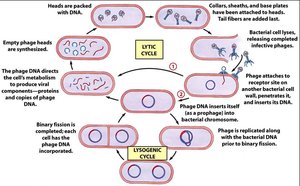
Phage conversion: Prophages can confer new pathogenic properties (e.g., toxin production) to bacteria.
Animal Virus Replication
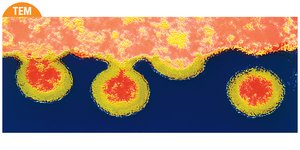
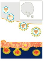
Animal viruses replicate in six steps: attachment, penetration, uncoating, replication, assembly, and release.
Attachment: Interaction with host cell membrane proteins
Penetration: Entry via fusion or endocytosis
Uncoating: Release of viral genome
Replication: Genome and protein synthesis
Assembly: Formation of new virions
Release: Budding (enveloped viruses) or cell lysis (naked viruses)


Persistent Infections
Some animal viruses cause persistent infections:
Chronic: Continuous release of virions, slow disease progression (e.g., HIV)
Latent: Intermittent flare-ups with periods of dormancy (e.g., herpesviruses)
Oncogenic Viruses
Oncoviruses can cause cancer by stimulating uncontrolled cell division or reducing cell death signals. Examples include human papilloma viruses (HPVs) and human T-lymphotropic viruses (HTLV).
Clinical Aspects of Viruses
Laboratory Growth of Viruses
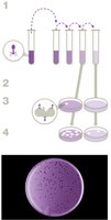
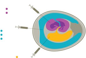
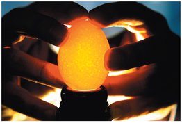
Viruses require host cells for laboratory cultivation. Bacteriophages are grown using plaque assays, where clear zones (plaques) indicate cell lysis. Animal viruses are grown in tissue culture, embryonated eggs, or live animal hosts.
Diagnostic Tests for Viral Detection
Diagnostic tests detect viral proteins, genetic material, or patient antibodies. Methods include:
Agglutination tests: Use antibodies or antigens linked to beads; positive result is bead clumping
ELISA: Detects antigens or antibodies via color change
Nucleic acid detection: PCR, sequencing, fluorescent probes

Antiviral Drugs
Antiviral drugs target various steps in viral replication, including attachment, penetration, uncoating, replication, assembly, and release. Most drugs limit infection rather than cure it. Vaccines are crucial for prevention.
Attachment inhibitors: Maraviroc (HIV), HRIG (rabies)
Penetration inhibitors: Enfuvirtide (HIV), docosanol (HHV-1)
Uncoating inhibitors: Amantadine (influenza)
Replication inhibitors: Nucleoside analogs (acyclovir, ribavirin), NRTIs (AZT)
Release inhibitors: Oseltamivir (Tamiflu), zanamivir (Relenza)
Interferons: Signal cells to limit viral entry and replication
Prions
Prions and Spongiform Encephalopathies
Prions are infectious proteins with no genetic material. They cause transmissible spongiform encephalopathies (TSEs), which can be inherited or acquired. Examples include Creuzfeldt-Jakob disease (CJD), Gerstmann-Straussler-Schienker syndrome, and fatal familial insomnia.
Transmission: Sporadic, inherited, iatrogenic, variant
Prion-like diseases: Alzheimer’s, Parkinson’s, ALS (misfolded proteins in brain)
Visual Summary