 뒤로
Viruses, Viroids, and Prions: Structure, Classification, and Pathogenicity
스터디 가이드 - 스마트 노트
 자료에 맞춘 맞춤형 노트, 핵심 정의, 예시, 맥락을 확장해 제공합니다.
General Characteristics of Viruses
Viruses vs. Bacteria
Viruses are fundamentally different from bacteria in their structure, replication, and metabolic capabilities. Unlike bacteria, viruses are obligate intracellular parasites that require a host cell for multiplication. They lack many cellular components found in bacteria, such as ribosomes and metabolic enzymes.
Viruses: Contain either DNA or RNA (never both), lack ATP-generating metabolism, ribosomes, and are sensitive to interferon but not antibiotics.
Bacteria: Possess both DNA and RNA, have ATP-generating metabolism, ribosomes, and are sensitive to antibiotics but not interferon.
Feature | Typical Bacteria | Rickettsias/Chlamydias | Viruses |
|---|---|---|---|
Intracellular Parasite | No | Yes | Yes |
Plasma Membrane | Yes | Yes | No |
Binary Fission | Yes | Yes | No |
Pass through Bacteriological Filters | No | Yes | Yes |
Possess Both DNA and RNA | Yes | Yes | No |
ATP-Generating Metabolism | Yes | No/Yes | No |
Ribosomes | Yes | Yes | No |
Sensitive to Antibiotics | Yes | Yes | No |
Sensitive to Interferon | No | No | Yes |
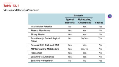
Example: Viruses cannot be treated with antibiotics, but some viral infections can be managed with interferons.
Viral Size
Viruses are much smaller than most bacteria, with sizes ranging from 20 to 1000 nm. Electron microscopy is required to visualize them. Some large viruses approach the size of small bacteria.
Smallest: Prions (~20 nm)
Largest: Ebola virus (~970 nm)
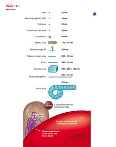
Example: Poliovirus is about 30 nm, while the Ebola virus is nearly 1000 nm.
Viral Structure
Virion and Nucleic Acid
A virion is a complete, infectious viral particle composed of nucleic acid and a protein coat. Viral genomes may be DNA or RNA, single- or double-stranded, linear or circular, and sometimes segmented.
Nucleic Acid: Encodes viral genes; can be single- or double-stranded, linear or circular.
Genome Size: Ranges from a few thousand to 250,000 nucleotides.
Example: Influenza virus has a segmented RNA genome.
Capsid and Envelope
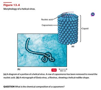
The capsid is a protein coat that protects viral nucleic acid. It is composed of subunits called capsomeres. Some viruses have an envelope derived from the host cell membrane, containing viral and host proteins, lipids, and carbohydrates. Spikes on the envelope aid in attachment and identification.
Nonenveloped viruses: Only have a capsid.
Enveloped viruses: Have a lipid/protein envelope surrounding the capsid.
Spikes: Glycoprotein projections used for attachment and identification.
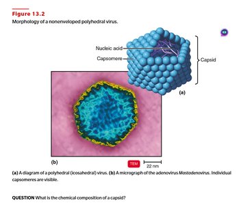
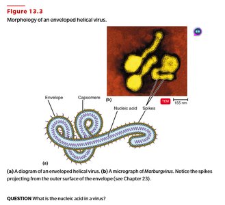
Example: Influenza virus is enveloped and has spikes that cause hemagglutination.
General Morphology
Viruses are classified by capsid architecture:
Helical: Rod-shaped, with nucleic acid inside a hollow cylinder (e.g., rabies, Ebola).
Polyhedral: Many-sided, often icosahedral (e.g., adenovirus, poliovirus).
Enveloped: Spherical, with envelope (e.g., Marburgvirus).
Complex: Complicated structures, often seen in bacteriophages (e.g., T-even phage).
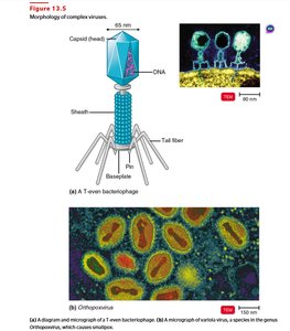
Example: Bacteriophages have a polyhedral head and helical tail.
Taxonomy of Viruses
Classification and Nomenclature
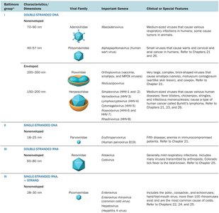
Viruses are classified based on their nucleic acid type and replication strategy. The Baltimore classification system divides viruses into seven groups. Family names end in -viridae, genus names in -virus, and order names in -virales.
Viral species: Group of viruses sharing genetic information and ecological niche.
Example: Family Herpesviridae, genus Simplexvirus, species human herpesvirus-2.
Baltimore Group | Characteristic/Dimension | Viral Family | Important Genes | Clinical Features |
|---|---|---|---|---|
I | Double-stranded DNA, nonenveloped | Mastadenovirus | Polyomavirus | Respiratory infections, tumors |
II | Single-stranded DNA, nonenveloped | Parvoviridae | Parvovirus | Fifth disease |
III | Double-stranded RNA, nonenveloped | Reoviridae | Rotavirus | Gastroenteritis |
IV | Single-stranded RNA, + strand | Picornaviridae | Enterovirus | Polio, colds |
V | Single-stranded RNA, - strand | Paramyxoviridae | Morbillivirus | Measles |
VI | Single-stranded RNA, + strand, produce DNA | Retroviridae | Lentivirus | HIV/AIDS |
VII | Double-stranded DNA, use reverse transcriptase, enveloped | Hepadnaviridae | Hepatitis B virus | Hepatitis, liver tumors |
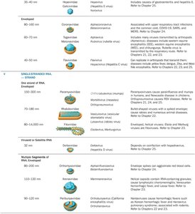
Example: Retroviruses (HIV) are classified as Group VI.
Isolation, Cultivation, and Identification of Viruses
Growing Bacteriophages
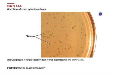
Bacteriophages are easily grown in bacterial cultures using the plaque method. Each plaque represents a single virus that has lysed bacterial cells.
Plaque-forming unit (PFU): Measurement of viral concentration based on plaque count.
Example: Plaques on E. coli lawn indicate bacteriophage activity.
Growing Animal Viruses
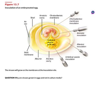
Animal viruses can be cultured in living animals, embryonated eggs, or cell cultures. Cell cultures are preferred for their convenience and homogeneity.
Embryonated eggs: Used for vaccine production; viral growth indicated by embryo death or lesions.
Cell cultures: Cells grown in media; viral infection causes cytopathic effects (CPE).
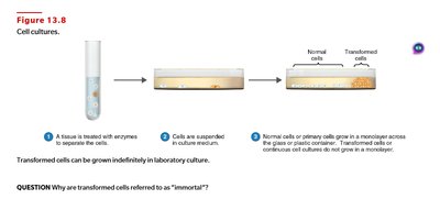
Example: HeLa cell line is a continuous cell culture used for viral research.
Viral Identification Techniques
Viruses are identified using serological methods (e.g., ELISA), observation of cytopathic effects, and molecular techniques such as PCR.
Serological tests: Detect viral antigens or antibodies.
PCR: Amplifies viral genetic material for identification.
Example: ELISA is used to detect HIV antigens in blood samples.
Viruses and Cancer
Oncogenic Viruses and Transformation
Some viruses can cause cancer by integrating their genetic material into the host genome, altering normal cell growth. Proto-oncogenes are normal genes that, when mutated, become oncogenes and trigger abnormal cell growth. Transformed cells exhibit uncontrolled growth and lack apoptosis.
Oncogenic viruses: DNA viruses (e.g., HPV, EBV, HBV) and retroviruses (e.g., HTLV-1, HTLV-2).
Oncolytic viruses: Used experimentally to treat cancer by selectively infecting and lysing tumor cells.
Example: HPV causes cervical cancer; retroviruses cause T cell leukemia.
Latent and Persistent Viral Infections
Latent vs. Persistent Infections
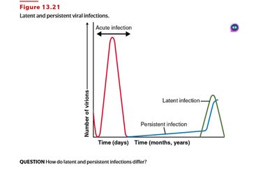
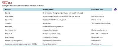
Viruses may remain dormant (latent) or continuously produce virions (persistent). Latent infections do not produce symptoms until reactivation, while persistent infections gradually worsen and are often fatal.
Latent: Cold sores (HSV), shingles (Varicella-zoster virus).
Persistent: HIV/AIDS, liver cancer (HBV, HCV), progressive encephalitis (measles).
Disease | Primary Effect | Causative Virus |
|---|---|---|
Cold sores | No symptoms during latency; viruses not usually released | HSV-1 and HSV-2 |
Leukemia | Increased white blood cell growth | HTLV-1 and 2 |
Shingles | Skin lesions | Varicellovirus (Herpesvirus) |
Cervical cancer | Increased cell growth | Human papillomavirus |
HIV/AIDS | Decreased CD4+ T cells | HIV-1 and 2 (Lentivirus) |
Liver cancer | Increased cell growth | Hepatitis B virus |
Progressive encephalitis | Rapid mental deterioration | Rubella virus |
Subacute sclerosing panencephalitis (SSPE) | Mental deterioration | Measles virus |
Example: Shingles occurs years after initial chickenpox infection.
Plant Viruses and Viroids
Plant Viruses
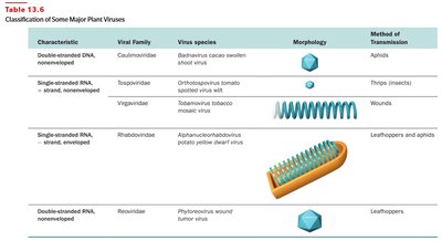
Plant viruses resemble animal viruses in structure and genome type. They cause diseases in crops and are transmitted via wounds or insect vectors. Some plant viruses can multiply in insect cells.
Characteristic | Viral Family | Virus Species | Morphology | Method of Transmission |
|---|---|---|---|---|
Double-stranded DNA, nonenveloped | Caulimoviridae | Badnavirus cacao swollen shoot virus | Icosahedral | Aphids |
Single-stranded RNA, nonenveloped | Virgaviridae | Tobamovirus tobacco mosaic virus | Helical | Wounds |
Single-stranded RNA, enveloped | Rhabdoviridae | Alphaphytorhabdovirus potato yellow dwarf virus | Enveloped helical | Leafhoppers and aphids |
Double-stranded RNA, nonenveloped | Reoviridae | Phytoreovirus wound tumor virus | Icosahedral | Leafhoppers |
Example: Tobacco mosaic virus is a helical plant virus transmitted by wounds.
Viroids and Virusoids
Viroids are short, naked RNA molecules that cause plant diseases. They lack a protein coat and are replicated by host RNA polymerase. Virusoids are viroids enclosed in a protein coat and require coinfection with a virus.
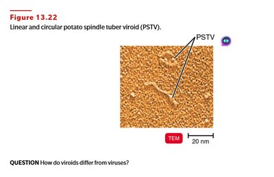
Example: Potato spindle tuber viroid causes crop losses.
Prions
Proteinaceous Infectious Particles
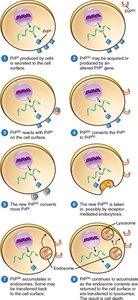
Prions are infectious proteins that cause neurological diseases by converting normal cellular prion protein (PrPC) into the abnormal form (PrPSc). Prion diseases are transmissible and involve degeneration of brain tissue.
Diseases: Scrapie (sheep), mad cow disease (cattle), Creutzfeldt-Jakob disease (humans).
Transmission: Ingestion, transplantation, contaminated instruments.
Example: Mad cow disease is transmitted to humans via contaminated meat.
Additional info: The notes include expanded explanations, definitions, and examples to ensure completeness and academic quality for microbiology students.
