 뒤로
뒤로Chapter 39: Assessment of Digestive and Gastrointestinal Function
스터디 가이드 - 스마트 노트
 자료에 맞춘 맞춤형 노트, 핵심 정의, 예시, 맥락을 확장해 제공합니다.
Chapter 39: Assessment of Digestive and Gastrointestinal Function
Anatomic and Physiologic Overview
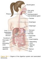
The gastrointestinal (GI) tract is a continuous tube approximately 7 to 7.9 meters (23 to 26 feet) in length, extending from the mouth to the anus. It includes the esophagus, stomach, small and large intestines, and rectum. The esophagus is located in the mediastinum, anterior to the spine and posterior to the trachea and heart, and passes through the diaphragm at the diaphragmatic hiatus.
Stomach: Located in the left upper abdomen, stores food, secretes digestive fluids, and propels chyme into the small intestine. It has four regions: cardia, fundus, body, and pylorus. The pyloric sphincter controls the opening to the small intestine.
Small Intestine: The longest segment, with three sections: duodenum, jejunum, and ileum. The ileocecal valve controls flow into the large intestine.
Large Intestine: Includes ascending, transverse, descending, and sigmoid colon, rectum, and anus. Regulated by internal and external anal sphincters.
Blood Supply and Innervation
The GI tract receives blood from arteries along the thoracic and abdominal aorta. The portal venous system collects blood from the GI tract and directs it to the liver. The GI tract is innervated by both sympathetic (inhibitory) and parasympathetic (stimulatory) nerves, which regulate motility, secretion, and sphincter control.
Sympathetic stimulation: Decreases gastric secretion and motility, constricts sphincters and blood vessels.
Parasympathetic stimulation: Increases peristalsis and secretory activities, relaxes sphincters.
External anal sphincter: Under conscious control.
Digestive Processes and Functions
Major Functions of the GI Tract
The digestive system breaks down food into molecular forms for absorption, absorbs nutrients into the bloodstream, and eliminates undigested material. Food is propelled through the GI tract and mixed with secretions that aid digestion and absorption.
Chewing and Swallowing
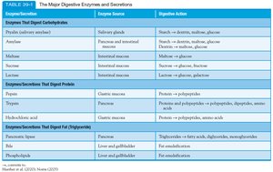
Digestion begins with chewing, which breaks food into small particles and mixes it with saliva. Salivation is triggered by the presence, sight, smell, or taste of food. Saliva contains ptyalin (salivary amylase) for starch digestion, as well as water and mucus for lubrication. Swallowing is a voluntary act regulated by the medulla oblongata, with the epiglottis preventing aspiration and esophageal peristalsis moving food to the stomach.
Gastric Function
The stomach stores and mixes food with highly acidic secretions (up to 2 L/day, pH as low as 1 due to hydrochloric acid). Gastric secretions break down food, destroy bacteria, and initiate protein digestion via pepsin. Intrinsic factor is necessary for vitamin B12 absorption; its absence leads to pernicious anemia.
Small Intestine Function
Digestion continues in the duodenum with secretions from the pancreas, liver, gallbladder, and intestinal glands. Pancreatic secretions are alkaline and contain enzymes such as trypsin, amylase, and lipase. Bile, produced by the liver and stored in the gallbladder, emulsifies fats. The small intestine's main function is nutrient absorption, with segmentation mixing contents and peristalsis propelling them forward.
Colonic Function
Waste material enters the colon within 4 hours after eating. Gut microbes assist in breaking down waste. Colonic secretions include electrolytes and mucus, with bicarbonate neutralizing bacterial end products. Slow peristalsis allows for water and electrolyte reabsorption, while strong waves propel contents after meals. Normal transit time is 24 to 48 hours.
Waste Products of Digestion
Feces consist of undigested food, inorganic materials, water, and bacteria (about 75% fluid, 25% solid). Brown color is due to bile breakdown; odor results from bacterial chemicals. Gases such as methane and hydrogen sulfide are absorbed or expelled. Elimination begins with rectal distention, triggering reflex contractions and relaxation of the internal anal sphincter. The external anal sphincter is under conscious control.
Gut Microbiome
The gut microbiota assist in waste breakdown, vitamin synthesis, and immune function. They protect against pathogens and regulate immune responses. Colonization begins shortly after birth and is established by age 2. Factors affecting microbiota include genetics, diet, hygiene, infection, aging, and medications. Disruption (e.g., by antibiotics) can lead to overgrowth of pathogenic species. Peyer patches play a role in immune defense.
Assessment of the Gastrointestinal System
Health History and Physical Assessment
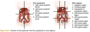
A focused GI assessment includes a complete history (abdominal pain, dyspepsia, gas, nausea, vomiting, diarrhea, constipation, jaundice, previous GI disease, nutrition, bowel habits, lifestyle) and a physical examination (mouth, abdomen, rectum). The abdomen is divided into four quadrants or nine regions for assessment.
Oral Cavity, Abdominal, and Rectal Examination
Oral cavity: Remove dentures for complete assessment; essential for detecting disorders such as cancer and diabetes.
Abdomen: Inspect for skin changes, auscultate before percussion and palpation, assess organ size and tenderness.
Rectum: External and internal examination for lumps, inflammation, tone, and irregularities.
Diagnostic Evaluation of GI Function
Laboratory Studies
Serum studies: CBC, metabolic panel, prothrombin time, triglycerides, liver function, amylase, lipase, tumor markers (CEA, CA 19-9, alpha-fetoprotein).
Stool tests: Consistency, color, occult blood, urobilinogen, fat, nitrogen, pathogens, and DNA tests. Guaiac-based fecal occult blood testing (gFOBT) and fecal immunochemical test (FIT) are used for cancer screening.
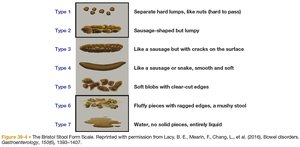
Bristol Stool Form Scale
The Bristol Stool Form Scale classifies stool into seven types, ranging from hard lumps (Type 1) to entirely liquid (Type 7). It is used to assess bowel function and diagnose disorders.
Breath Tests
Hydrogen breath test: Evaluates carbohydrate absorption and bacterial overgrowth.
Urea breath test: Detects Helicobacter pylori infection, a cause of peptic ulcer disease.
Genetic Testing
Genetic risk assessment is used for conditions such as gastric cancer, lactose deficiency, inflammatory bowel disease, and colon cancer. Lynch syndrome is the most common hereditary cause of colorectal cancer.
Imaging Studies
Ultrasonography: Noninvasive, no radiation, immediate results.
CT and MRI: Provide cross-sectional images for diagnosis of inflammatory, neoplastic, and structural conditions.
Upper and lower GI studies: Use contrast agents (e.g., barium) to visualize the GI tract.
Endoscopic Procedures
Esophagogastroduodenoscopy (EGD): Visualizes the esophagus, stomach, and duodenum; allows for biopsy and cytology.
Colonoscopy: Direct inspection of the large intestine for cancer screening and diagnosis. Preparation includes bowel cleansing and dietary restrictions.
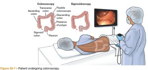
Anoscopy, proctoscopy, sigmoidoscopy: Examine the anus, rectum, and sigmoid colon for chronic diarrhea, bleeding, and other disorders.
Small bowel studies: Capsule endoscopy and double-balloon enteroscopy visualize the small intestine.
Manometry and electrophysiologic studies: Assess GI motility disorders by measuring intraluminal pressures.
Gastric analysis and pH monitoring: Diagnose secretory disorders and reflux disease.
Laparoscopy: Minimally invasive evaluation of abdominal organs, often for diagnosis of peritoneal disease or chronic pain.
Gerontologic Considerations
Age-Related Changes in the GI System
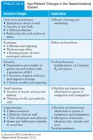
Aging has minimal direct effect on most GI functions, but older adults are more likely to experience disorders such as anorexia, nausea, vomiting, indigestion, and changes in bowel habits. Normal physiologic changes with aging include alterations in oral cavity, esophagus, stomach, small intestine, and large intestine function.
Sample Questions and Answers
Question 1
Which of the following is NOT a function of the stomach?
A. Storing food
B. Secreting digestive fluids
C. Propelling chyme into the small intestine
D. Absorbing nutrients
E. Mixing food with secretions
Answer: D. Absorbing nutrients Rationale: Nutrient absorption primarily occurs in the small intestine, not the stomach.
Question 2
Is the following statement true or false? Aging has minimal direct effect on most GI functions.
Answer: True Rationale: Most GI functions remain relatively stable with age, though older adults may experience common GI disorders.
Question 3
Which of the following GI disorders is most commonly associated with Lynch syndrome?
A. Gastric cancer
B. Lactose deficiency
C. Inflammatory bowel disease
D. Colon cancer
E. Portal hypertension
Answer: D. Colon cancer Rationale: Lynch syndrome is the most common cause of hereditary colorectal cancer.
Additional info:
Table of major digestive enzymes and their sources/actions is included in image_2.
Table of age-related GI changes and implications is included in image_3.
Bristol Stool Form Scale (image_5) is a clinical tool for stool classification.
