 뒤로
뒤로Chapter 40: Management of Patients with Oral and Esophageal Disorders
스터디 가이드 - 스마트 노트
 자료에 맞춘 맞춤형 노트, 핵심 정의, 예시, 맥락을 확장해 제공합니다.
Chapter 40: Management of Patients with Oral and Esophageal Disorders
Oral Health and Periodontal Disease
Oral health is essential for both physical and psychological well-being. Periodontal disease, including gingivitis and periodontitis, is a major concern due to its prevalence and impact on overall health.
Gingivitis: Inflammation of the gums, preventable with consistent oral hygiene and regular dental cleanings.
Periodontitis: Advanced gum disease involving inflammation of gums, soft tissue, and bone structures. Symptoms include soreness, bleeding gums, halitosis, pain with chewing, changes in facial appearance, and tooth loss.
Prevalence: 42% of the U.S. population has periodontitis; 8% have severe periodontitis. Higher rates in males, Mexican Americans, non-Hispanic Black Americans, smokers, those with uncontrolled diabetes, and those living below the poverty line.
Dental Plaque and Caries
Dental plaque and caries are common oral health issues caused by biofilms and poor hygiene.
Dental Plaque: Hardened biofilm of microorganisms that cannot be removed by routine brushing.
Dental Caries: Demineralization of dental hard tissues, leading to tooth decay or cavities. Contributing factors include genetics, high-sugar diet, smoking, alcohol, and poor oral hygiene.
Prevalence: Untreated caries affect 10% of adolescents, 21% of adults, and 13% of older adults.
Periapical Abscess
A periapical abscess is a collection of pus at the apex of a tooth, resulting from infection.
Acute Abscess: Arises from infection due to caries, trauma, or failed root canal.
Chronic Abscess: Slowly progressive, may form a granuloma and often discovered via x-ray.
Symptoms: Dull, gnawing pain, swelling, temperature sensitivity, tooth mobility, fever, malaise, difficulty opening mouth.
Oral Health and Chronic Disease
There is a strong link between oral health and chronic diseases such as diabetes, cardiovascular disease, osteoporosis, and others.
Mechanism: Gram-negative bacteria in periodontal disease trigger systemic inflammation, increasing risk for cardiovascular disease.
Recommendations: Prevention and treatment of periodontitis, routine questioning during cardiology workups, collaboration between cardiologists and dentists.
Disorders of the Jaw
Temporomandibular Disorders (TMD)
TMDs affect the jaw joint and muscles, causing pain and limited motion.
Types: Disorders of masticatory muscles (myalgia), joint pain (arthralgia), displaced disc, bone destruction.
Symptoms: Jaw pain, restricted motion, locking, changes in bite, clicking/popping sounds, headaches, earaches, dizziness.
Diagnosis: Based on pain and motion limitations; MRI for severe cases.
Treatment: Noninvasive therapies (self-care, stress reduction, physical therapy, integrative health interventions), analgesics, oral appliances, psychiatric management for comorbidities.
Jaw Disorders Requiring Surgery
Structural abnormalities or trauma may require surgical intervention.
Procedures: Jaw wiring, reconstruction, bone grafting, fixation techniques.
Complications: Infection, malunion, hardware failure, missed injuries.
Nursing Management: Liquid/soft diet, oral care, medication adherence, abstaining from smoking/alcohol, regular follow-up.
Disorders of the Salivary Glands
Salivary Gland Function and Disorders
Salivary glands produce saliva for lubrication, protection, and digestion. Disorders include inflammation, stones, and neoplasms.
Parotitis: Inflammation of the parotid gland, caused by infection, dehydration, or autoimmune disorders. Symptoms include fever, swelling, pain, difficulty swallowing.
Sialadenitis: Inflammation of salivary glands, often due to stones (sialolithiasis). Managed with massage, antibiotics, sialagogues, heat, hydration.
Neoplasms: Tumors in salivary glands, diagnosed by biopsy and treated with surgery, radiation, or chemotherapy.
Cancer of the Oral Cavity and Pharynx
Overview and Risk Factors
Cancers of the mouth and throat are curable if detected early. Risk factors include tobacco, alcohol, HPV infection, and previous head and neck cancer.
Pathophysiology: Most are squamous cell carcinomas; HPV is linked to 70% of oropharyngeal cancers.
Symptoms: Early stages have few symptoms; later stages show sores, patches, ulcers, difficulty chewing/swallowing, weight loss, neck mass.
Diagnosis: Oral exam, lymph node assessment, imaging (PET-CT, MRI), endoscopy, biopsy, HPV testing.
Treatment: HPV vaccine, surgical resection, radiation, chemotherapy, reconstructive surgery.
Nursing Management: Nutritional assessment, communication support, airway maintenance, graft viability assessment, tracheostomy care.
Nursing Management of Oral Cavity Disorders
Promoting Mouth Care and Comfort
Oral care before and during cancer treatment reduces complications such as mucositis.
Protocols: Multiagent oral care combinations; avoid chlorhexidine and sucralfate in stomatitis.
Ensuring Adequate Food and Fluid Intake
Nutrition is vital for recovery and maintaining energy levels. Enteral feedings may be necessary.
Factors: Weight, age, activity, oral discomfort, dysphagia, taste changes.
Recommendations: Involve a registered dietitian, monitor calorie intake, use supplements as needed.
Supporting a Positive Self-Image
Patients may experience changes in body image due to illness or treatment. Support and counseling are important.
Strategies: Encourage verbalization, realistic discussion, support groups, psychiatric referral.
Preventing Infection
Patients are at increased risk for infection due to leukopenia, malnutrition, and diabetes.
Monitoring: Frequent lab checks, temperature monitoring, aseptic technique, wound care.
Educating Patients About Self-Care
Education on mouth care, nutrition, infection prevention, and feeding techniques is essential for patient independence.
Methods: Demonstrate techniques, facilitate return demonstration, provide written instructions.
Continuing and Transitional Care
Ongoing care and follow-up are necessary for recovery and health maintenance.
Home Care: Teach suctioning, odor control, humidification, monitor for complications, reinforce health promotion.
Neck Dissection
Types and Complications
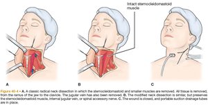
Neck dissection is performed to remove cancerous lymph nodes and surrounding tissues. There are several types with varying degrees of tissue removal.
Radical Neck Dissection: Removal of all cervical lymph nodes, sternocleidomastoid muscle, internal jugular vein, and spinal accessory nerve.
Modified Radical/Selective Dissection: Preserves some nonlymphatic structures or lymph node groups.
Complications: Shoulder dysfunction, poor cosmesis, nerve injury.
Reconstructive Techniques
Reconstruction may involve cutaneous, myocutaneous, or microvascular free flaps from various body sites.
Sites: Scapula, radial forearm, anterolateral thigh.
Disorders of the Esophagus
Overview
The esophagus is a muscular tube that transports food from the mouth to the stomach. Disorders include motility issues, hernias, diverticula, perforation, foreign bodies, chemical burns, GERD, Barrett esophagus, benign tumors, and cancer.
Dysphagia: Most common symptom, ranging from discomfort to acute pain.
Esophageal Motility Disorders
Motility disorders such as achalasia and distal esophageal spasm affect swallowing and esophageal function.
Achalasia: Ineffective peristalsis and failure of LES to relax, leading to esophageal dilation.
Symptoms: Dysphagia, regurgitation, chest pain, heartburn.
Diagnosis: X-ray, barium swallow, CT, endoscopy, high-resolution manometry.
Treatment: Slow eating, fluid intake, PPIs, smooth muscle relaxants, botulinum toxin, pneumatic dilation.
Hiatal Hernia
Hiatal hernia occurs when part of the stomach moves up into the thorax through an enlarged diaphragm opening.
Types: Sliding (type I) and paraesophageal.
Symptoms: Heartburn, regurgitation, dysphagia, GERD symptoms, chronic cough.
Diagnosis: Endoscopy, manometry, pH monitoring, barium swallow.
Treatment: Small meals, upright posture, PPIs, surgical repair if needed.
Diverticulum
Esophageal diverticula are outpouchings of the esophageal wall, most commonly Zenker diverticulum.
Symptoms: Dysphagia, neck fullness, belching, regurgitation, halitosis.
Diagnosis: Barium swallow, manometry.
Treatment: Endoscopic or open surgery, diet progression post-surgery.
Perforation
Esophageal perforation is a surgical emergency requiring immediate diagnosis and treatment.
Causes: Iatrogenic, spontaneous, trauma, malignancy.
Symptoms: Severe pain, dysphagia, infection, hypotension.
Treatment: NPO, IV fluids, antibiotics, surgical repair, nutritional support.
Foreign Bodies and Chemical Burns
Swallowed foreign bodies and chemical burns can cause injury or obstruction and require prompt management.
Symptoms: Pain, dysphagia, dyspnea, severe burns, breathing difficulties.
Treatment: Removal, shock management, NPO, IV fluids, antibiotics, nutritional support.
Gastroesophageal Reflux Disease (GERD)
GERD is the backflow of gastric contents into the esophagus, causing symptoms and mucosal injury.
Causes: Incompetent LES, hiatal hernia, motility disorder, lifestyle factors.
Symptoms: Heartburn, regurgitation, dyspepsia, dysphagia, esophagitis, complications (ulcerations, strictures, cancer).
Diagnosis: History, pH monitoring, manometry, endoscopy.
Treatment: Lifestyle modifications, PPIs, surgical intervention if needed.
Barrett Esophagus
Barrett esophagus is a precancerous condition where the esophageal lining changes, increasing risk for adenocarcinoma.
Symptoms: Frequent heartburn, peptic ulcer symptoms, esophageal stricture.
Diagnosis: Endoscopy, biopsy.
Treatment: Endoscopic ablation, surveillance, PPIs.
Benign and Malignant Tumors of the Esophagus
Benign tumors are rare; malignant tumors include adenocarcinoma and squamous cell carcinoma.
Symptoms: Dysphagia, pain, regurgitation, weight loss.
Diagnosis: Imaging, biopsy, laboratory tests.
Treatment: Surgery, radiation, chemotherapy, palliative care.
Nursing Management: Nutritional support, pulmonary care, family education, monitoring for complications.
Enteral Nutrition and Tube Feeding
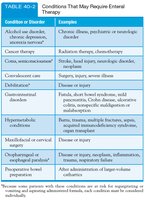
Indications for Enteral Therapy
Enteral therapy is indicated for patients unable to meet nutritional needs orally due to various conditions.
Condition or Disorder | Examples |
|---|---|
Alcohol use disorder, chronic depression, anorexia nervosa | Chronic illness, psychiatric or neurologic disease |
Cancer therapy | Radiation therapy, chemotherapy |
Cardiovascular event | Stroke, heart injury, vascular disease |
Disordered eating | Trauma, sports severe illness |
Diabetes | Diabetic food issues, metabolic syndrome, renal compromise, cardiac insufficiency |
Hypermetabolic syndrome | Burns, trauma, multiple illnesses, sepsis, metabolic disturbances |
Modified oral or cervical | Disease or injury |
Oropharyngeal or esophageal impairment | Disease or injury, neoplasms, inflammation |
Preoperative bowel preparation | Abdominal surgery, large-volume resection |
Administration and Management of Tube Feedings
Tube feedings are used when oral intake is inadequate or impossible, and the GI tract is functional.
Types: Nasoduodenal, nasojejunal, gastrostomy, jejunostomy tubes.
Formula Selection: Based on GI status, nutritional needs, osmolality, caloric density, fiber content.
Administration Methods: Large-bore NG tubes for short-term, small-bore nasoenteric tubes for up to 6 weeks, gastrostomy/jejunostomy for long-term.
Monitoring: Flow rate, fluid intake, feeding tolerance, tube patency, infection prevention.
Complications and Nursing Diagnoses
Potential complications include infection, skin breakdown, tube dislodgment, GI bleeding, and obstruction.
Nursing Diagnoses: Impaired nutrition, risk for infection, impaired skin integrity, disturbed body image.
Goals: Achieve nutritional requirements, prevent infection, maintain skin integrity, adjust to body image changes, prevent complications.
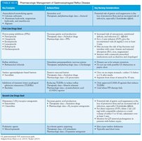
Summary Table: Pharmacologic Management of GERD
Key Symptoms | Drug Class | Actions | Key Nursing Considerations |
|---|---|---|---|
Pyrosis, regurgitation, dyspepsia, dysphagia, odynophagia, hypersalivation, esophagitis | Proton pump inhibitors (PPIs), H2-receptor antagonists, antacids | Decrease gastric acid secretion, neutralize acid | Monitor for side effects, educate on proper use, assess symptom relief |
Persistent symptoms | Prokinetic agents, mucosal protectants | Increase gastric emptying, protect mucosa | Monitor for adverse effects, assess GI function |
Refractory symptoms | Second-line drugs, surgical options | Target underlying causes, surgical correction | Monitor post-surgical recovery, educate on lifestyle changes |
