 뒤로
뒤로Anatomical Language and Microscopy: Study Guide for Anatomy & Physiology
스터디 가이드 - 스마트 노트
 자료에 맞춘 맞춤형 노트, 핵심 정의, 예시, 맥락을 확장해 제공합니다.
Chapter 1: Anatomical Language
Anatomical Position and Body Organization
The anatomical position is the standard reference for describing locations and directions on the human body. Understanding this position and related terminology is essential for accurate communication in anatomy and physiology.
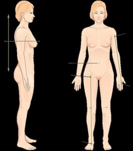
Anatomical Position: The body stands upright, facing forward, arms at the sides with palms facing forward, feet together.
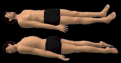
Prone Position: Lying face down.
Supine Position: Lying face up.
Body Organization: The human body is organized into regions, cavities, and systems for study and clinical reference.
Directional Terms
Directional terms are used to describe the location of one body part relative to another.
Superior (cranial): Above or toward the head.
Inferior (caudal): Below or toward the feet.
Anterior (ventral): Toward the front of the body.
Posterior (dorsal): Toward the back of the body.
Medial: Toward the midline.
Lateral: Away from the midline.
Proximal: Closer to the point of attachment.
Distal: Farther from the point of attachment.
Superficial (external): Nearer to the surface.
Deep (internal): Farther from the surface.
Body Planes and Sections
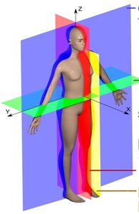
Body planes are imaginary lines used to divide the body for anatomical study and medical imaging.
Sagittal Plane: Divides the body into right and left portions.
Midsagittal Plane: Divides the body into equal right and left halves.
Parasagittal Plane: Divides the body into unequal right and left portions.
Frontal (Coronal) Plane: Divides the body into anterior and posterior portions.
Transverse (Horizontal) Plane: Divides the body into superior and inferior portions.
Oblique Plane: Cuts at an angle other than horizontal or vertical.
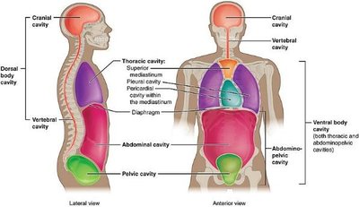
Body Cavities and Membranes
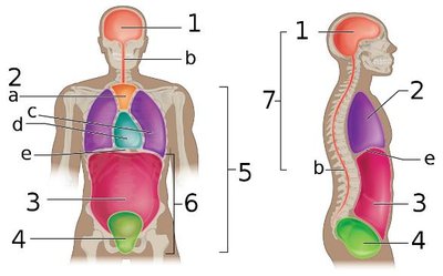
The body contains several cavities that house organs and are lined by membranes for protection and lubrication.
Dorsal Cavity: Contains the cranial cavity (brain) and vertebral cavity (spinal cord).
Ventral Cavity: Contains the thoracic cavity (heart, lungs), abdominopelvic cavity (digestive, urinary, reproductive organs).
Serous Membranes: Thin tissues that line cavities and cover organs. Includes pleura (lungs), pericardium (heart), and peritoneum (abdominal organs).
Membrane Layers: Visceral (covers organs), Parietal (lines cavity walls), with a serous cavity between them containing lubricating fluid.
Abdominopelvic Quadrants and Regions
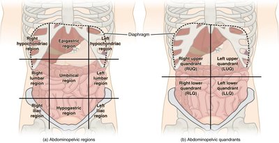
The abdominopelvic cavity is divided into quadrants and regions for clinical reference.
Quadrants: Right Upper (RUQ), Right Lower (RLQ), Left Upper (LUQ), Left Lower (LLQ).
Regions: Epigastric, Left/Right Hypochondriac, Umbilical, Left/Right Lumbar, Hypogastric, Left/Right Iliac.
Clinical Application: Used to localize pain, masses, or organ pathology.
Regional Terminology
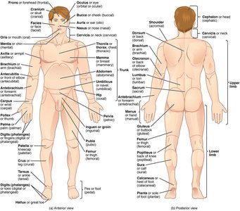
Regional terms specify areas of the body for anatomical study and clinical practice.
Axial Region: Head (cephalic), neck (cervical), trunk (thoracic, abdominal, pelvic).
Appendicular Region: Upper extremity (arm, forearm, wrist, hand), lower extremity (thigh, leg, ankle, foot).
Examples: Brachial (arm), Femoral (thigh), Patellar (kneecap), Plantar (sole of foot).
Organ Systems Overview
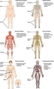
The human body is organized into organ systems, each with specific functions and major organs.
Integumentary System: Skin, hair, nails; protection, vitamin D synthesis.
Skeletal System: Bones, cartilage; support, movement, protection.
Muscular System: Muscles; movement, heat production.
Nervous System: Brain, spinal cord, nerves; control, communication.
Endocrine System: Glands; hormone secretion, regulation.
Cardiovascular System: Heart, blood vessels; transport, regulation.
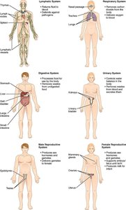
Lymphatic System: Lymph nodes, vessels; immunity, fluid balance.
Respiratory System: Lungs, airways; gas exchange.
Digestive System: Stomach, intestines; nutrient breakdown and absorption.
Urinary System: Kidneys, bladder; waste removal, fluid balance.
Reproductive System: Ovaries/testes; reproduction.
Chapter 2: Use of the Microscope
Microscope Parts and Functions
The compound light microscope is an essential tool for studying cells and tissues in anatomy and physiology. Understanding its parts and proper use is critical for laboratory work.
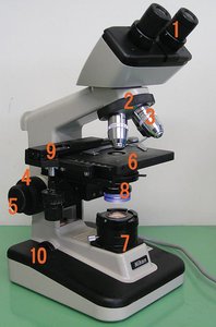
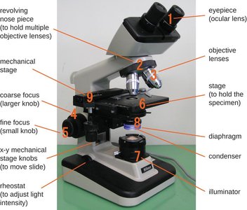
Ocular Lens (Eyepiece): Magnifies the image, usually 10x.
Objective Lenses: Provide varying magnifications (4x, 10x, 40x, 100x).
Revolving Nosepiece (Turret): Holds and rotates objective lenses.
Mechanical Stage: Holds the slide and allows precise movement.
Stage Clips: Secure the slide.
Coarse Focus Knob: Large adjustments for focusing (low magnification).
Fine Focus Knob: Small adjustments for sharp focus (high magnification).
Condenser: Focuses light onto the specimen.
Iris Diaphragm: Controls light intensity.
Light Source (Illuminator): Provides illumination.
Base and Arm: Support and carry the microscope.
Microscope Operation and Magnification
Proper use and calculation of magnification are fundamental skills in microscopy.
Carrying: Always use two hands (one on the arm, one on the base).
Cleaning: Use lens paper and solution for lenses.
Magnification Formula:
Total Magnification:
Field of View: The visible area through the microscope decreases as magnification increases.
Depth of Field: The thickness of the specimen in focus; decreases with higher magnification.
Resolution: The ability to distinguish two points as separate.
Measuring Field of View
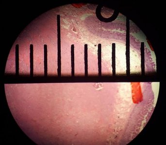
Field of view can be measured using a slide with a metric ruler. Calculation is required for higher magnifications.
Measurement: Align ruler marks across the diameter of the field.
Calculation Formula:
Microscopy Glossary
Binocular: Microscope with two eyepieces.
Brightfield Microscope: Produces a dark image on a bright background.
Coarse/Fine Focus: Knobs for large/small adjustments.
Condenser: Focuses light.
Ocular Lens: Eyepiece.
Objective Lens: Closest to specimen.
Resolution: Ability to distinguish points.
Stage: Platform for slides.
Total Magnification: Product of ocular and objective magnification.
Additional info:
All content is based on the objectives and terminology required for introductory Anatomy & Physiology courses, including regional, directional, and systemic anatomy, as well as microscopy fundamentals.
Tables referenced in the workbook (e.g., organ systems, quadrants, regions) are described in text; students should refer to their lab manual for completion and practice.
