 뒤로
뒤로Anatomical Terminology and Body Organization
스터디 가이드 - 스마트 노트
 자료에 맞춘 맞춤형 노트, 핵심 정의, 예시, 맥락을 확장해 제공합니다.
Anatomical Terminology and Body Organization
Introduction to Anatomical Terminology
Anatomical terminology provides a standardized language for describing the structure, position, and relationships of parts of the human body. This foundational vocabulary is essential for clear communication in anatomy and physiology.
Standard Anatomical Position
Definition and Importance
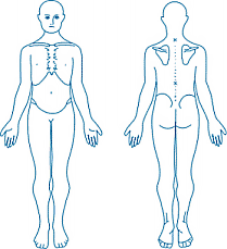
The anatomical position is the reference posture used to describe the locations and relationships of body parts. In this position, the individual stands erect, faces forward, arms at the sides, palms facing forward, and feet parallel and directed forward. This standardization eliminates ambiguity when describing anatomical structures.
Directional Terms
Basic Directional Terms
Directional terms are used to describe the location of one body part relative to another. These terms are essential for accurately communicating anatomical relationships.
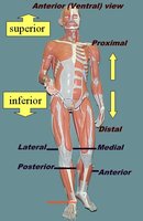
Superior: Toward the head or upper part of a structure (e.g., the nose is superior to the chin).
Inferior: Away from the head or toward the lower part of a structure (e.g., the knees are inferior to the hips).
Anterior (Ventral): Toward the front of the body (e.g., the chest is anterior to the spine).
Posterior (Dorsal): Toward the back of the body (e.g., the spine is posterior to the heart).
Medial: Toward the midline of the body (e.g., the nose is medial to the eyes).
Lateral: Away from the midline of the body (e.g., the arms are lateral to the chest).
Proximal: Closer to the point of attachment or origin (e.g., the elbow is proximal to the wrist).
Distal: Farther from the point of attachment or origin (e.g., the fingers are distal to the elbow).
Superficial: Closer to the surface of the body (e.g., the skin is superficial to the muscles).
Deep: Farther from the surface of the body (e.g., the bones are deep to the muscles).
Additional Directional Terms
Ipsilateral: On the same side of the body (e.g., the right hand and right foot are ipsilateral).
Contralateral: On the opposite side of the body (e.g., the right hand and left foot are contralateral).
Summary Table of Directional Terms
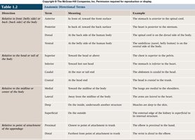
Direction | Term | Meaning | Example |
|---|---|---|---|
Front/Back | Anterior (Ventral) | Toward the front | The stomach is anterior to the spinal cord. |
Front/Back | Posterior (Dorsal) | Toward the back | The spinal cord is posterior to the heart. |
Up/Down | Superior | Toward the head | The chest is superior to the pelvis. |
Up/Down | Inferior | Toward the feet | The stomach is inferior to the heart. |
Midline | Medial | Toward the midline | The lungs are medial to the shoulders. |
Midline | Lateral | Away from the midline | The arms are lateral to the heart. |
Attachment | Proximal | Closer to attachment | The elbow is proximal to the wrist. |
Attachment | Distal | Farther from attachment | The wrist is distal to the elbow. |
Surface | Superficial | Closer to surface | The skin is superficial to the muscles. |
Surface | Deep | Farther from surface | The bone is deep to the muscle. |
Body Planes and Sections
Major Body Planes
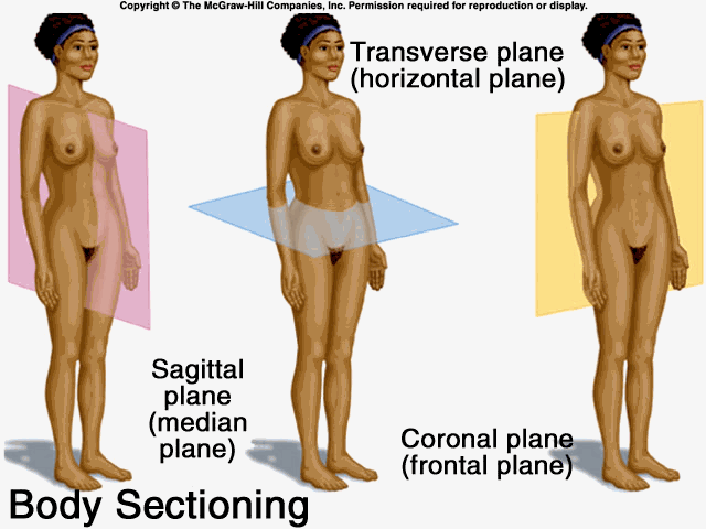
Body planes are imaginary flat surfaces that divide the body into sections. They are used to describe views and cuts in anatomical studies and medical imaging.
Frontal (Coronal) Plane: Divides the body into anterior (front) and posterior (back) portions.
Sagittal Plane: Divides the body into right and left portions. A midsagittal plane divides the body into equal right and left halves, while a parasagittal plane divides it into unequal portions.
Transverse (Horizontal) Plane: Divides the body into superior (upper) and inferior (lower) portions; also called a cross-section.
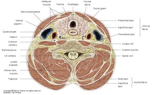
Example: Transverse Section at C6 Vertebra
A transverse section at the level of the C6 vertebra provides a cross-sectional view of the neck, revealing anatomical relationships among muscles, blood vessels, and other structures.
Regional Anatomical Terms
Anterior and Posterior Body Regions
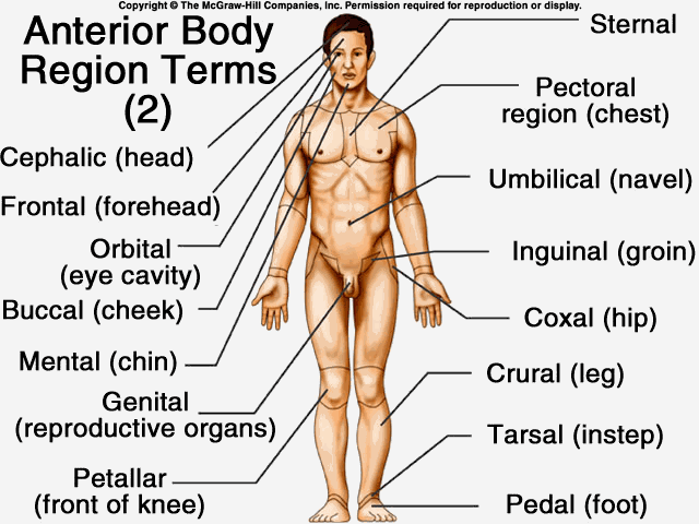
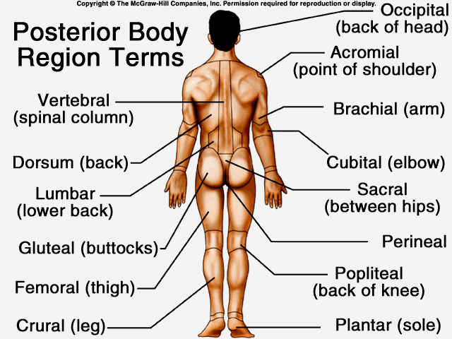
Regional terms specify distinct areas of the body, aiding in precise anatomical description. These terms are often used in clinical and anatomical contexts to localize injuries, diseases, or procedures.
Cephalic: Head
Cervical: Neck
Pectoral: Chest
Umbilical: Navel
Inguinal: Groin
Coxal: Hip
Crural: Leg
Tarsal: Ankle
Pedal: Foot
Occipital: Back of head
Vertebral: Spinal column
Gluteal: Buttocks
Femoral: Thigh
Plantar: Sole of foot
Summary Table of Body Regions
Regional Name | Description | Regional Name | Description |
|---|---|---|---|
Abdominal | Region inferior to the thorax and superior to the hip bones | Manual | Hand |
Antebrachial | Forearm | Mental | Chin |
Axillary | Armpit | Oral | Mouth |
Brachial | Arm | Palmar | Palm of the hand |
Buccal | Cheek | Patellar | Kneecap |
Carpal | Wrist | Pedal | Foot |
Cephalic | Head | Perineal | Region between anus and external reproductive organs |
Body Cavities
Major Body Cavities and Their Subdivisions
The body contains internal spaces called body cavities that house and protect organs. The two main cavities are the dorsal and ventral cavities, each with further subdivisions.
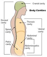
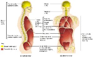
Dorsal Cavity: Includes the cranial cavity (brain) and spinal cavity (spinal cord).
Ventral Cavity: Includes the thoracic cavity (lungs and heart) and abdominopelvic cavity (digestive, urinary, and reproductive organs).
Abdominopelvic Cavity
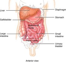
The abdominopelvic cavity is subdivided into the abdominal cavity (containing digestive organs) and the pelvic cavity (containing urinary bladder, reproductive organs, and rectum).
Thoracic Cavity
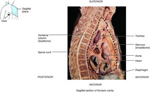
The thoracic cavity is located above the diaphragm and contains the lungs and heart, protected by the rib cage.
Summary Table: Body Cavities and Membranes
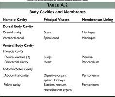
Name of Cavity | Principal Viscera | Membranous Lining |
|---|---|---|
Cranial cavity | Brain | Meninges |
Vertebral canal | Spinal cord | Meninges |
Pleural cavities | Lungs | Pleurae |
Pericardial cavity | Heart | Pericardium |
Abdominal cavity | Digestive organs, spleen, kidneys | Peritoneum |
Pelvic cavity | Bladder, rectum, reproductive organs | Peritoneum |
Body Membranes
Serous Membranes
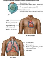
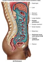
Serous membranes line body cavities that do not open to the exterior and cover the organs within these cavities. Each serous membrane has a parietal layer (lining the cavity wall) and a visceral layer (covering the organ).
Pericardium: Surrounds the heart.
Pleura: Surrounds the lungs.
Peritoneum: Surrounds abdominal organs.
Abdominal Regions and Quadrants
Four Abdominal Quadrants
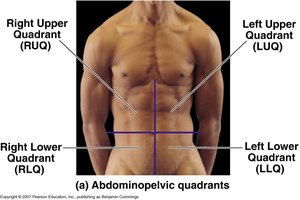
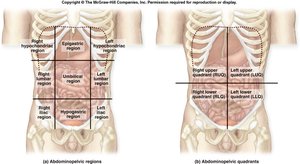
The abdomen is divided into four quadrants for clinical reference: right upper (RUQ), left upper (LUQ), right lower (RLQ), and left lower (LLQ). These quadrants help localize pain, injuries, or disease.
Nine Abdominal Regions
For more precise localization, the abdomen is also divided into nine regions: right and left hypochondriac, right and left lumbar, right and left inguinal, epigastric, umbilical, and hypogastric (pubic) regions.
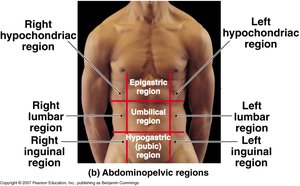
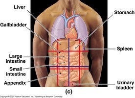
Abdominal Regions and Quadrants with Underlying Organs
These divisions are used to describe the locations of underlying organs and to guide diagnosis and treatment.
Summary
Understanding anatomical terminology, body planes, regions, and cavities is essential for accurately describing the structure and organization of the human body. Mastery of these concepts forms the foundation for further study in anatomy and physiology.
