뒤로
뒤로Anatomical Terminology and Body Organization: Foundations for Anatomy & Physiology
스터디 가이드 - 스마트 노트
 자료에 맞춘 맞춤형 노트, 핵심 정의, 예시, 맥락을 확장해 제공합니다.
Anatomical Terminology
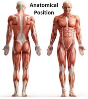
Anatomical Position
The anatomical position is the standard reference posture used to describe the location and relation of body parts. In this position, the body stands upright, facing forward, with arms at the sides and palms facing forward. All anatomical terminology assumes the body is in this position, regardless of its actual posture during examination or injury.
Directional Terms
Overview of Directional Terms
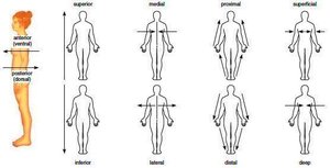
Directional terms are used by medical professionals and anatomists to describe the locations of structures relative to other structures or locations in the body. These terms provide clarity and precision in communication.
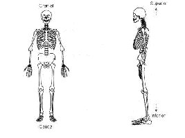
Superior (Cranial or Cephalic) and Inferior (Caudal)
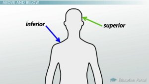
Superior: Toward the head end or upper part of the body; above.
Inferior: Away from the head end or toward the lower part of a structure or the body; below.
Example: The head is superior to the abdomen; the knees are inferior to the hips.
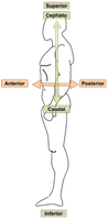
Anterior (Ventral) and Posterior (Dorsal)
Anterior (Ventral): Toward or at the front of the body; in front of.
Posterior (Dorsal): Toward or at the back of the body; behind.
Example: The sternum is anterior to the heart; the spine is posterior to the lungs.
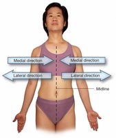
Medial, Lateral, and Intermediate
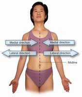
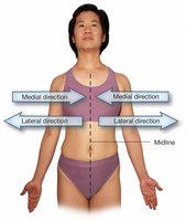
Medial: Toward or at the midline of the body; on the inner side of.
Lateral: Away from the midline of the body; on the outer side of.
Intermediate: Between a more medial and a more lateral structure.
Example: The nose is medial to the eyes; the arms are lateral to the chest; the collarbone is intermediate between the breastbone and shoulder.
Proximal and Distal
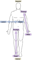
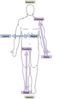
Proximal: Closer to the origin of the body part or the point of attachment of a limb to the body trunk.
Distal: Farther from the origin of a body part or the point of attachment of a limb to the body trunk.
Example: The elbow is proximal to the wrist; the fingers are distal to the elbow.
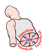
Superficial (External) and Deep (Internal)
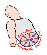
Superficial (External): Toward or at the body surface.
Deep (Internal): Away from the body surface; more internal.
Example: The skin is superficial to the muscles; the lungs are deep to the rib cage.
Regional Terms
Anterior Body Landmarks
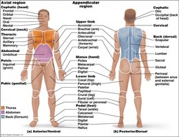
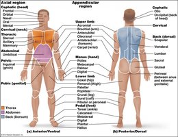
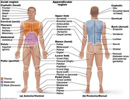
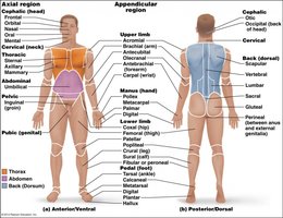
Regional terms specify visible landmarks on the anterior (front) and posterior (back) surfaces of the body. These terms are essential for accurately describing locations of injuries, incisions, or anatomical features.
Abdominal: Anterior body trunk inferior to ribs
Antebrachial: Forearm
Antecubital: Anterior surface of elbow
Axillary: Armpit
Brachial: Arm
Buccal: Cheek area
Carpal: Wrist
Cervical: Neck region
Crural: Anterior lower leg between knee and ankle
Digital: Fingers, toes
Femoral: Thigh (applies to both anterior and posterior)
Fibular: Lateral part of leg
Frontal: Forehead
Nasal: Nose area
Oral: Mouth
Orbital: Eye area
Patellar: Anterior knee
Pelvic: Area overlying the pelvis anteriorly
Pubic: Genital region
Sternal: Breastbone area
Tarsal: Ankle region
Thoracic: Area between neck and abdomen
Umbilical: Navel
Posterior Body Landmarks
Cephalic: Head
Femoral: Thigh
Lumbar: Area of back between ribs and hips; the loin
Occipital: Posterior surface of head or base of skull
Olecranon: Bony eminence of ulna (elbow)
Popliteal: Posterior knee area
Scapular: Shoulder blade region
Sural: Muscular swelling of posterior leg below knee
Vertebral: Area of spinal column
Body Planes and Sections
Major Body Planes
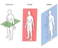
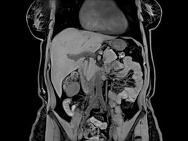
Body planes are imaginary lines used to divide the body into sections for anatomical study and medical imaging. The three primary planes are:
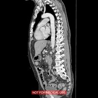
Sagittal Plane: Divides the body into left and right parts. If exactly in the midline, it is called the midsagittal or median plane; if offset, it is a parasagittal plane.
Frontal (Coronal) Plane: Divides the body into anterior (front) and posterior (back) parts.
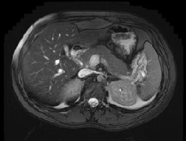
Transverse (Horizontal) Plane: Divides the body into superior (upper) and inferior (lower) parts.
Body Cavities
Dorsal and Ventral Body Cavities
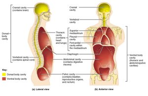
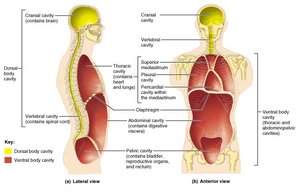
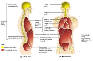
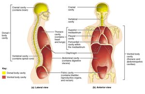
The body contains internal cavities that protect organs and allow for changes in organ shape and size. The two main cavities are:
Dorsal Body Cavity: Includes the cranial cavity (houses the brain) and the spinal cavity (encloses the spinal cord).
Ventral Body Cavity: Larger and contains the thoracic cavity (chest), mediastinum (central compartment of thoracic cavity), and abdominopelvic cavity (abdominal and pelvic organs).
Other Body Cavities
Oral and Digestive Cavity: Mouth and continuous digestive organs.
Nasal Cavity: Within and posterior to the nose; part of the respiratory system.
Orbital Cavities: House the eyes.
Middle Ear Cavities: Medial to the eardrums; contain bones for hearing.
Abdominopelvic Regions and Quadrants
Quadrants of the Abdominopelvic Cavity
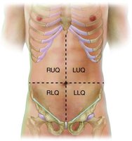
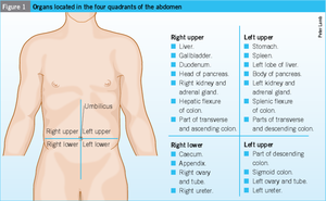
The abdominopelvic cavity is divided into four quadrants for clinical reference:
Right Upper Quadrant (RUQ)
Left Upper Quadrant (LUQ)
Right Lower Quadrant (RLQ)
Left Lower Quadrant (LLQ)
Nine Abdominopelvic Regions
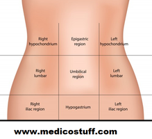
For more precise anatomical study, the abdominopelvic cavity is divided into nine regions:
Umbilical Region: Center, surrounding the navel
Epigastric Region: Superior to the umbilical region
Hypogastric (Pubic) Region: Inferior to the umbilical region
Left and Right Iliac (Inguinal) Regions: Lateral to the hypogastric region
Left and Right Lumbar Regions: Lateral to the umbilical region
Left and Right Hypochondriac Regions: Flank the epigastric region and contain the lower ribs