 뒤로
뒤로Anatomy & Physiology 1 Lab Exam 1 Study Guidance
스터디 가이드 - 스마트 노트
 자료에 맞춘 맞춤형 노트, 핵심 정의, 예시, 맥락을 확장해 제공합니다.
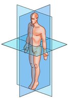
Q1. Label the picture of the planes of the body.
Background
Topic: Anatomical Planes
This question tests your understanding of the three major anatomical planes used to describe locations and directions in the human body. These planes are essential for communicating about body structure and function in anatomy.
Key Terms:
Sagittal Plane: Divides the body into left and right portions.
Frontal (Coronal) Plane: Divides the body into anterior (front) and posterior (back) portions.
Transverse (Horizontal) Plane: Divides the body into superior (upper) and inferior (lower) portions.
Step-by-Step Guidance
Observe the image and identify the three planes intersecting the body.
Recall the definitions of sagittal, frontal (coronal), and transverse planes.
Match each plane in the image to its definition based on how it divides the body.
Label each plane accordingly, but do not write the final labels yet.
Try solving on your own before revealing the answer!
Final Answer:
The plane dividing the body into left and right is the sagittal plane.
The plane dividing the body into front and back is the frontal (coronal) plane.
The plane dividing the body into upper and lower parts is the transverse (horizontal) plane.
These planes are fundamental for describing anatomical locations and movements.
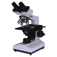
Q2. Label the parts of the microscope.
Background
Topic: Light Microscope Anatomy
This question assesses your knowledge of the main structural components of a compound light microscope, which is essential for proper use and understanding of microscopy in anatomy labs.
Key Terms:
Ocular lens (eyepiece): The lens you look through, usually 10x magnification.
Objective lenses: Multiple lenses with different magnifications (e.g., 4x, 10x, 40x, 100x).
Stage: The platform where the slide is placed.
Coarse and fine focus knobs: Used to focus the image.
Light source: Illuminates the specimen.
Arm and base: Support the microscope structure.
Step-by-Step Guidance
Examine the image and identify the main visible parts of the microscope.
Recall the function and location of each part listed above.
Match each part in the image to its correct anatomical term.
Prepare to label each part, but do not write the final labels yet.
Try solving on your own before revealing the answer!
Final Answer:
Ocular lens (eyepiece): Top part you look through.
Objective lenses: Rotating nosepiece with multiple lenses.
Stage: Flat platform for slides.
Coarse and fine focus knobs: Large and small knobs on the side.
Light source: Located at the base, shines light upward.
Arm: Curved structure connecting base and head.
Base: Bottom support of the microscope.
Knowing these parts is essential for using the microscope effectively in lab settings.
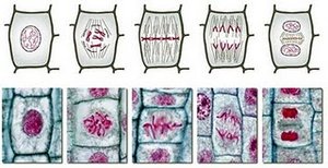
Q3. Label the stages of mitosis.
Background
Topic: Mitosis (Cell Division)
This question tests your ability to recognize and label the stages of mitosis, a process of cell division that results in two genetically identical daughter cells. Understanding mitosis is fundamental in anatomy and physiology for learning about growth, repair, and cellular reproduction.
Key Terms:
Prophase: Chromatin condenses, spindle forms, nuclear envelope breaks down.
Metaphase: Chromosomes align at the metaphase plate.
Anaphase: Sister chromatids separate and move to opposite poles.
Telophase: Chromosomes decondense, nuclear envelope reforms.
Cytokinesis: Division of the cytoplasm, resulting in two cells.
Step-by-Step Guidance
Observe the sequence of images showing the progression of mitosis.
Recall the defining features of each mitotic stage listed above.
Match each image to the correct stage based on visible characteristics (e.g., chromosome alignment, separation).
Prepare to label each stage, but do not write the final labels yet.
Try solving on your own before revealing the answer!
Final Answer:
First image: Prophase
Second image: Metaphase
Third image: Anaphase
Fourth image: Telophase
Fifth image: Cytokinesis
Each stage has distinct features, such as chromosome condensation, alignment, separation, and cell division.
