 뒤로
뒤로Anatomy & Physiology Exam 1 Study Guide – Step-by-Step Guidance
스터디 가이드 - 스마트 노트
 자료에 맞춘 맞춤형 노트, 핵심 정의, 예시, 맥락을 확장해 제공합니다.
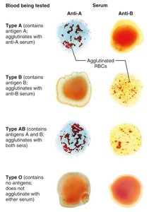
Q97. Be able to tell me which blood type given this image for each Matching
Background
Topic: Blood Typing and Agglutination
This question tests your understanding of blood types (A, B, AB, O) and how agglutination reactions with anti-A and anti-B sera are used to identify blood types.
Key Terms and Concepts:
Agglutination: Clumping of red blood cells when antibodies bind to antigens.
Antigen: Protein on the surface of RBCs (A or B).
Serum: Contains antibodies (anti-A or anti-B).
Blood Types: A, B, AB, O (based on presence/absence of A and B antigens).
Step-by-Step Guidance
Examine the image showing blood samples mixed with anti-A and anti-B sera. Look for agglutination (clumping) in each sample.
Recall: Agglutination with anti-A serum means the blood has A antigens; agglutination with anti-B serum means the blood has B antigens.
Type A: Agglutinates with anti-A, not anti-B. Type B: Agglutinates with anti-B, not anti-A. Type AB: Agglutinates with both. Type O: No agglutination with either.
Match each blood sample in the image to its blood type based on the agglutination pattern.
Set up your answer by listing the blood types and describing the agglutination results for each.
Try solving on your own before revealing the answer!
Final Answer:
Type A: Agglutinates with anti-A serum only.
Type B: Agglutinates with anti-B serum only.
Type AB: Agglutinates with both anti-A and anti-B sera.
Type O: Does not agglutinate with either serum.
The image shows the agglutination patterns for each blood type, helping you visually identify them based on the reaction with the sera.
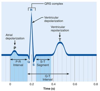
Q135. Be able to label and know details for PQRS wave
Background
Topic: Electrocardiogram (ECG/EKG) Waves
This question tests your knowledge of the components of an ECG tracing, including the P wave, QRS complex, T wave, and intervals/segments.
Key Terms and Concepts:
P wave: Represents atrial depolarization.
QRS complex: Represents ventricular depolarization.
T wave: Represents ventricular repolarization.
PR interval, ST segment, QT interval: Important intervals/segments for cardiac function.
Step-by-Step Guidance
Study the ECG image and identify the labeled waves: P, Q, R, S, T.
Recall what each wave represents: P (atrial depolarization), QRS (ventricular depolarization), T (ventricular repolarization).
Locate the intervals and segments: PR interval, ST segment, QT interval.
Understand the timing and sequence: P wave occurs first, followed by QRS, then T wave.
Set up your answer by labeling each part and describing its physiological significance.
Try solving on your own before revealing the answer!
Final Answer:
P wave: Atrial depolarization.
QRS complex: Ventricular depolarization.
T wave: Ventricular repolarization.
PR interval: Time from atrial to ventricular depolarization.
ST segment: Time between ventricular depolarization and repolarization.
QT interval: Duration of ventricular depolarization and repolarization.
The image helps you visualize and label each wave and interval, reinforcing their physiological roles.
