뒤로
뒤로Anatomy and Physiology of the Heart
스터디 가이드 - 스마트 노트
 자료에 맞춘 맞춤형 노트, 핵심 정의, 예시, 맥락을 확장해 제공합니다.
The Heart: Structure and Function
Overview of Heart Location and Orientation
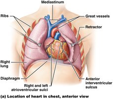
The heart is a muscular organ located in the thoracic cavity, specifically within the mediastinum, between the lungs. It is slightly tilted so that the apex points to the left hip and the base is directed toward the right shoulder. The heart is protected by the rib cage and rests on the diaphragm.
Mediastinum: Central compartment of the thoracic cavity containing the heart, great vessels, and other structures.
Diaphragm: Muscular partition separating the thoracic and abdominal cavities; the heart sits atop the diaphragm.
Great vessels: Major arteries and veins entering and leaving the heart, including the aorta, pulmonary arteries, and veins.
Layers of the Heart Wall and Pericardium
Structure of the Heart Wall
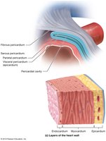
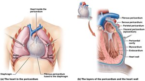
The heart wall is composed of three distinct layers, each with specialized functions. Surrounding the heart is the pericardium, a double-walled sac that provides protection and reduces friction during heartbeats.
Epicardium: The outermost layer, also known as the visceral pericardium; provides a protective layer and contains blood vessels.
Myocardium: The thick, muscular middle layer responsible for the contractile force of the heart.
Endocardium: The innermost layer lining the heart chambers and valves; composed of simple squamous epithelium.
Pericardium: Consists of the fibrous pericardium (outer tough layer) and serous pericardium (parietal and visceral layers) with a lubricating pericardial cavity in between.
External Anatomy of the Heart
Major External Features
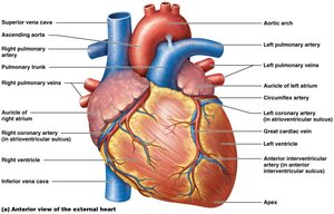
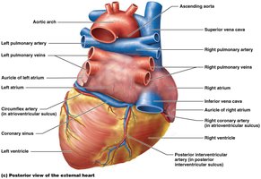
The heart's external anatomy includes several key structures visible from anterior and posterior views. These features are important for understanding the flow of blood and the attachment of major vessels.
Atria: The two upper chambers (right and left atria) that receive blood returning to the heart.
Ventricles: The two lower chambers (right and left ventricles) that pump blood out of the heart.
Major vessels: Include the aorta, pulmonary trunk, pulmonary veins, and venae cavae.
Sulci: Grooves on the heart surface marking the boundaries between chambers and containing coronary vessels (e.g., anterior and posterior interventricular sulci, atrioventricular sulcus).
Internal Anatomy of the Heart
Heart Chambers and Valves
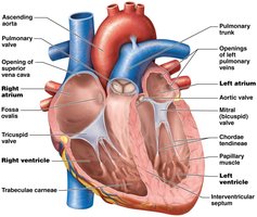
The heart contains four chambers: two atria and two ventricles. Valves ensure unidirectional blood flow and prevent backflow during the cardiac cycle.
Right Atrium: Receives deoxygenated blood from the body via the superior and inferior vena cava.
Right Ventricle: Pumps deoxygenated blood to the lungs via the pulmonary trunk.
Left Atrium: Receives oxygenated blood from the lungs via the pulmonary veins.
Left Ventricle: Pumps oxygenated blood to the body via the aorta.
Valves: Tricuspid (right AV), bicuspid/mitral (left AV), pulmonary semilunar, and aortic semilunar valves.
Circuits of Blood Flow
Pulmonary and Systemic Circuits
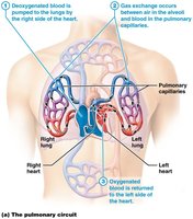
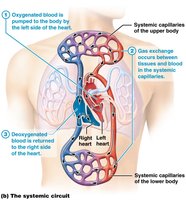
The heart pumps blood through two main circuits: the pulmonary circuit (to and from the lungs) and the systemic circuit (to and from the rest of the body). Each circuit serves a distinct function in oxygenating blood and delivering nutrients.
Pulmonary Circuit: Carries deoxygenated blood from the right ventricle to the lungs and returns oxygenated blood to the left atrium.
Systemic Circuit: Carries oxygenated blood from the left ventricle to the body and returns deoxygenated blood to the right atrium.
Ventricular Structure and Function
Comparison of Right and Left Ventricles
The right and left ventricles differ in wall thickness and shape, reflecting their distinct roles in pumping blood to the lungs and the systemic circulation, respectively.
Ventricle | Wall Thickness | Chamber Shape |
|---|---|---|
Right Ventricle | Thin | Crescent-shaped |
Left Ventricle | Thick | Circular |
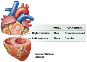
Example: The left ventricle must generate higher pressure to pump blood throughout the body, requiring a thicker muscular wall compared to the right ventricle, which only pumps blood to the nearby lungs.
Coronary Circulation
Arterial and Venous Supply of the Heart
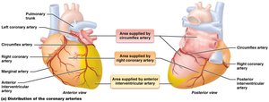
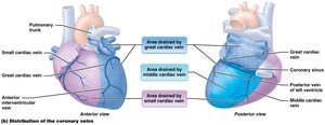
The heart muscle (myocardium) receives its own blood supply through the coronary arteries and is drained by the coronary veins. This circulation is essential for delivering oxygen and nutrients to cardiac tissue.
Coronary Arteries: Branch from the aorta and supply oxygenated blood to the heart muscle. Major branches include the right and left coronary arteries, circumflex artery, and anterior/posterior interventricular arteries.
Coronary Veins: Collect deoxygenated blood from the myocardium and return it to the right atrium via the coronary sinus. Major veins include the great, middle, and small cardiac veins.
Coronary Artery | Area Supplied |
|---|---|
Right Coronary Artery | Right atrium, right ventricle, part of left ventricle |
Left Coronary Artery | Left atrium, left ventricle, interventricular septum |
Circumflex Artery | Lateral and posterior walls of left ventricle |
Anterior Interventricular Artery | Anterior walls of both ventricles |
Coronary Vein | Area Drained |
|---|---|
Great Cardiac Vein | Anterior surface of the heart |
Middle Cardiac Vein | Posterior interventricular sulcus |
Small Cardiac Vein | Right margin of the heart |
Cardiac Muscle Tissue
Structure and Function of Cardiac Muscle
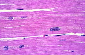
Cardiac muscle tissue is specialized for continuous, rhythmic contraction. It is composed of branching cells connected by intercalated discs, which allow for rapid transmission of electrical impulses and coordinated contraction.
Striations: Alternating light and dark bands due to the arrangement of actin and myosin filaments.
Intercalated Discs: Specialized junctions that connect cardiac muscle cells, facilitating synchronized contraction and efficient heart function.
Involuntary Control: Cardiac muscle contracts without conscious control, regulated by the autonomic nervous system and intrinsic conduction system.