 뒤로
뒤로Anatomy & Physiology Study Guide: Foundations and Skeletal System
스터디 가이드 - 스마트 노트
 자료에 맞춘 맞춤형 노트, 핵심 정의, 예시, 맥락을 확장해 제공합니다.
How to Succeed in Your Anatomy and Physiology Course
Reading Strategies: The SQ3R Method
The SQ3R method is a proven approach for reading textbooks effectively, especially in fast-paced courses like Anatomy & Physiology. It stands for Survey, Question, Read, Recite, and Review.
Survey: Skim the chapter to get an overview of the main topics and structure.
Question: Formulate questions based on headings and learning outcomes.
Read: Read actively, seeking answers to your questions.
Recite: Summarize key points aloud or in writing.
Review: Go over the material again to reinforce learning.
Example: Before reading about the skeletal system, ask "What are the main divisions of the skeleton?" and "How do bones support movement?"
Using the Textbook and Associated Materials
The textbook is organized into modules, each beginning with learning outcomes. It includes concept boosts, study boosts, and numerous questions to reinforce learning. Figures are essential for visualizing anatomical structures and processes.
Modules: Each module covers a specific topic.
Learning Outcomes: Guide your study by focusing on what you should know.
Figures: Use figures to understand spatial relationships and functions.
Overview of Anatomy & Physiology
Characteristics of Living Organisms
Living organisms share several fundamental characteristics that distinguish them from non-living matter.
Organization: Structured arrangement from molecules to organ systems.
Metabolism: All chemical reactions occurring in the body.
Responsiveness: Ability to detect and respond to stimuli.
Growth: Increase in size and number of cells.
Reproduction: Production of new cells or organisms.
Movement: Both internal and external motion.
Structural Levels of Organization
The human body is organized into hierarchical levels, each building upon the previous.
Chemical Level: Atoms and molecules.
Cellular Level: Cells, the basic unit of life.
Tissue Level: Groups of similar cells performing a function.
Organ Level: Structures composed of multiple tissue types.
Organ System Level: Groups of organs working together.
Organism Level: The complete living being.
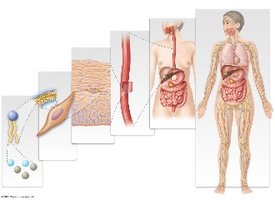
Example: The heart is an organ composed of muscle tissue, connective tissue, and nervous tissue, functioning within the cardiovascular system.
Approaches to Anatomy
Systemic Anatomy: Study of body systems (e.g., cardiovascular, skeletal).
Regional Anatomy: Study of specific regions (e.g., head, neck).
Gross Anatomy: Structures visible to the naked eye.
Microscopic Anatomy: Structures requiring magnification (e.g., histology).
Example: In this course, the systemic approach is primarily used.
The Language of Anatomy & Physiology
Organ Systems
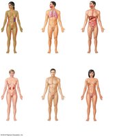
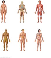
Organ systems are groups of organs that work together to perform major functions. Understanding their classification is foundational.
Examples: Digestive system, respiratory system, nervous system, etc.
Classification: Based on function and structure.
Medical Terminology: Roots, Prefixes, Suffixes
Medical terms are constructed from word roots, prefixes, suffixes, and combining vowels.
Word Root: The base of a term (e.g., cardi for heart).
Prefix: Added to the beginning (e.g., hyper- means above normal).
Suffix: Added to the end (e.g., -itis means inflammation).
Combining Vowel: Often "o" to connect roots (e.g., oste/o for bone).
Example: Cardiology = cardi (heart) + ology (study of).
Anatomical Position and Directional Terms
The anatomical position is the standard reference for describing locations and directions in the body.
Anatomical Position: Standing upright, facing forward, arms at sides, palms forward.
Directional Terms: Used to describe relative positions (e.g., superior, inferior, anterior, posterior).
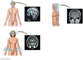
Planes of Section
Planes of section are imaginary lines used to divide the body for anatomical study.
Sagittal Plane: Divides body into left and right.
Frontal (Coronal) Plane: Divides body into anterior and posterior.
Transverse Plane: Divides body into superior and inferior.
Oblique Plane: Cuts at an angle.
Example: MRI images are often taken in sagittal, coronal, or transverse planes.
The Organization of the Human Body
Body Cavities
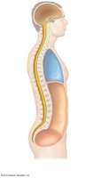
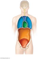
Body cavities house and protect internal organs. They are divided into dorsal and ventral cavities.
Dorsal Body Cavity: Contains the cranial and spinal cavities.
Ventral Body Cavity: Contains the thoracic and abdominopelvic cavities.
Thoracic Cavity: Subdivided into pleural, pericardial, and mediastinum.
Abdominopelvic Cavity: Subdivided into abdominal and pelvic cavities.
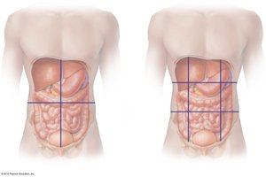
Abdominal Quadrants and Regions
The abdominopelvic cavity is divided for clinical and anatomical reference.
Four Quadrants: Right upper, left upper, right lower, left lower.
Nine Regions: Right/left hypochondriac, epigastric, right/left lumbar, umbilical, right/left iliac, hypogastric.
Serous Membranes
Serous membranes line body cavities and cover organs, reducing friction.
Two Layers: Parietal (lines cavity) and visceral (covers organ).
Serous Fluid: Lubricates surfaces.
Pleural Membranes: Surround lungs.
Pericardial Membranes: Surround heart.
Peritoneal Membranes: Surround abdominal organs.
Retroperitoneal: Organs located behind the peritoneum (e.g., kidneys).
Medical Imaging in Anatomy & Physiology
Imaging Techniques
Medical imaging is essential for visualizing internal structures.
X-Rays: Use radiation to view dense structures like bones.
CT Scans: Use X-rays and computer processing for cross-sectional images.
MRI: Uses magnetic fields and radio waves for detailed images of soft tissues.
Overview of the Skeletal System
Axial and Appendicular Skeleton
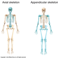
The skeleton is divided into two main parts: axial and appendicular. The axial skeleton includes the skull, vertebral column, and thoracic cage, while the appendicular skeleton includes the limbs and girdles.
Axial Skeleton: Skull, vertebral column, thoracic cage.
Appendicular Skeleton: Pectoral girdle, upper limbs, pelvic girdle, lower limbs.
Number of Bones: A typical adult has 206 bones.
Bone Markings
Bones have markings that serve as sites for muscle attachment, passage of nerves and blood vessels, and articulation with other bones. These markings are classified as depressions, openings, or projections.
Type | Example | Function |
|---|---|---|
Depression | Fossa | Muscle attachment or articulation |
Opening | Foramen | Passage for nerves/vessels |
Projection | Process | Muscle attachment |
Example: The foramen magnum in the skull allows passage of the spinal cord.
Additional info:
Some content was inferred for completeness, such as the classification of bone markings and the summary of medical imaging techniques.
