 뒤로
뒤로Anatomy & Physiology Study Guide: Introduction, Histology, Integumentary, and Skeletal Systems
스터디 가이드 - 스마트 노트
 자료에 맞춘 맞춤형 노트, 핵심 정의, 예시, 맥락을 확장해 제공합니다.
Introduction to Anatomy and Physiology
Fields of Anatomy and Physiology
Anatomy and physiology are foundational sciences for understanding the human body. Anatomy is the study of structure, often referred to as the "naming of parts," while physiology is the study of function, focusing on "how does it work."
Anatomy: Structure of body parts
Physiology: Function of body parts
Types/Fields of Anatomy and Physiology
There are several specialized fields within anatomy and physiology:
Systemic anatomy: Study of individual organ systems
Regional anatomy: Study by specific body regions
Surface anatomy: Study of surface markings
Gross anatomy: Structures visible to the naked eye
Microscopic anatomy: Structures seen under a microscope
Histology: Study of tissues
Cytology: Study of cells
Cell physiology: Study of cell function
Neuroanatomy: Study of nervous system structures
Digestive physiology: Study of digestive functions
Characteristics of Life
All living things share certain characteristics:
Made of cells
Show metabolism (anabolic and catabolic processes)
Can grow (increase in cell number or size)
Perform excretion (removal of waste)
Respond to stimuli
Show movement
Can reproduce
Levels of Organization
The human body is organized from least to most complex:
Chemical level
Cellular level
Tissue level
Organ level
Organ system level
Organism level
Organ Systems of the Human Body
There are eleven organ systems, each with specific components and functions:
Integumentary: Protection, vitamin D production, water retention, temperature regulation
Skeletal: Support, protection, movement, blood cell production, calcium storage
Muscular: Movement, control of openings, heat generation
Nervous: Regulation, sensation, movement, higher functions
Endocrine: Regulation via hormones
Cardiovascular: Transport of blood, nutrients, wastes
Lymphatic: Fluid return, immunity
Respiratory: Gas exchange, acid-base balance
Digestive: Digestion, absorption, waste removal, balance maintenance
Urinary: Waste removal, balance maintenance, blood cell production
Reproductive: Gamete production, hormone secretion, fetal development
Anatomical Position and Bilateral Symmetry
The anatomical position is a standard reference: standing upright, feet shoulder-width apart, arms at sides, head and palms facing forward. Bilateral symmetry means the body can be divided into mirror-image halves along one plane.
Body Region and Directional Terms
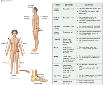
Body region terms (e.g., mental for chin) and directional terms (e.g., anterior, posterior, superior, inferior, medial, lateral, superficial, deep) are essential for describing locations and relationships in the body.
Anterior: Toward the front
Posterior: Toward the back
Superior: Toward the head
Inferior: Toward the tail
Medial: Closer to the midline
Lateral: Farther from the midline
Proximal: Closer to the point of attachment
Distal: Farther from the point of attachment
Superficial: Closer to the surface
Deep: Farther below the surface
Sections and Planes
Body planes are used to describe anatomical cuts:
Sagittal: Right and left sections
Midsagittal: Equal right and left halves
Parasagittal: Unequal right and left sections
Frontal (coronal): Anterior and posterior sections
Transverse (horizontal): Superior and inferior sections
Oblique: Angled sections
Abdominopelvic Quadrants and Regions
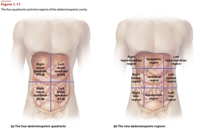
The abdomen is divided into quadrants and regions for clinical and anatomical reference:
Quadrants: Right upper (RUQ), Left upper (LUQ), Right lower (RLQ), Left lower (LLQ)
Regions: Right/left hypochondriac, epigastric, right/left lumbar, umbilical, right/left iliac, hypogastric
Body Cavities
The body contains several cavities, each housing specific organs:
Cranial cavity: Brain
Vertebral (spinal) cavity: Spinal cord
Thoracic cavity: Heart and lungs
Pleural cavities: Lungs
Mediastinum: Heart
Abdominopelvic cavity: Digestive organs
Serous Membranes
Serous membranes are two-layered membranes with fluid between layers, reducing friction and holding organs in place. The visceral layer contacts the organ, while the parietal layer attaches to surrounding structures. Types include pleural (lungs), pericardium (heart), and peritoneum (abdominal organs).
Homeostasis and Regulation
Homeostasis is the tendency to maintain a stable internal environment. Each variable has a set point and a normal range. Regulation occurs via autoregulation (intrinsic) and extrinsic regulation (nervous and endocrine systems).
Feedback Loops
Feedback loops maintain homeostasis:
Negative feedback: Opposes change, returns to set point (most common)
Positive feedback: Magnifies change, moves away from set point (rare, e.g., blood clotting, labor)
Dynamic Equilibrium and Disease
The body is in a state of dynamic equilibrium, always changing within a normal range. Disease occurs when homeostasis cannot be maintained.
Medical Imaging Techniques
Common imaging techniques include:
X-Ray: Uses radiation to view dense structures
MRI: Uses magnetic fields for detailed images
CT (CAT) Scan: Uses X-rays for cross-sectional images
PET Scan: Shows metabolic activity
Ultrasound: Uses sound waves for real-time imaging
Histology: The Study of Tissues
Four Basic Types of Tissue
The human body contains four basic tissue types:
Epithelium: Covers surfaces, forms glands, has polarity, cellularity, regeneration, and secretion
Muscle: Responsible for movement and force
Connective: Provides support, storage, cushioning, transport
Nervous: Generates electrical signals, controls functions
Cell Junctions
Cell junctions include:
Desmosomes: Resist mechanical stress
Gap junctions: Allow communication between cells
Tight junctions: Make spaces impermeable
Glandular Epithelium
Glands are classified as:
Endocrine: Release hormones into blood
Exocrine: Release products via ducts
Modes of exocrine secretion:
Merocrine: Release only product
Apocrine: Release product plus part of cell
Holocrine: Release product plus whole cell
Extracellular Matrix and Protein Fibers
The extracellular matrix (ECM) contains protein fibers:
Collagen: Thick, strong
Reticular: Thin, supportive
Elastic: Stretchy, spiral-like
Connective Tissue Cell Types
Connective tissues contain various cell types, each with specific functions.
Muscle Tissue Types
Muscle tissue is classified as skeletal, cardiac, or smooth, each with unique functions and locations.
Nervous Tissue
Nervous tissue consists of neurons (signal transmission) and neuroglial cells (support).
Histology Terms
Regeneration: Replacement of damaged tissue
Fibrosis: Formation of scar tissue
Scar: Result of fibrosis
Cancer: Uncontrolled cell growth
Germ Layers
Three primary germ layers give rise to adult tissues:
Ectoderm: Skin, nervous tissue
Mesoderm: Muscle, connective tissue
Endoderm: Digestive and respiratory linings
The Integumentary System
Functions and Layers of Skin
The integumentary system protects, regulates temperature, and produces vitamin D. Skin has multiple layers:
Epidermis: Outer layer, contains various cell types
Dermis: Inner layer, contains accessory structures
Epidermal Cell Types and Pigments
Cell types include keratinocytes, melanocytes, and others. Pigments include melanin, carotene, and hemoglobin.
Skin Growth and Types
Skin grows via cell division; thick and thin skin differ in structure and location.
Dermis Structure and Accessory Structures
The dermis contains connective tissue, glands, hair follicles, and nails.
Cleavage Lines and Skin Cancer
Cleavage lines are important in surgery. Types of skin cancer vary in severity.
Hair, Nails, and Glands
Hair follicles and nails grow from specialized structures. Glands include sweat and sebaceous glands.
Body Temperature Regulation
The skin regulates temperature via sweat and blood flow.
Burns and Complications
Burns are classified by depth; major complications include fluid loss and infection. The "rule of nines" estimates burn area.
Common Skin Disorders
Psoriasis, Acne, Jaundice, Cyanosis, Albinism, Erythema, Pallor, Decubitus ulcers, Stretch marks, Callus
The Skeletal System
Functions and Bone Shapes
The skeletal system supports, protects, enables movement, produces blood cells, and stores minerals. Bones are classified by shape: long, short, flat, irregular, sesamoid.
Bone Anatomy and Matrix
Long bones have distinct gross and microscopic anatomy. The bony matrix is composed of collagen and minerals.
Compact vs. Spongy Bone
Compact bone is dense; spongy bone is porous and found at bone ends.
Bone Marrow
Red marrow: Blood cell production; Yellow marrow: Fat storage.
Bone Cells
Types include osteoblasts (build), osteoclasts (break down), osteocytes (maintain).
Bone Formation and Growth
Intramembranous and endochondral ossification are processes of bone formation. Bone growth occurs at growth plates.
Bone Remodeling and Repair
Remodeling is ongoing; repair follows fracture with specific steps.
Calcium Regulation
Calcitonin and PTH regulate calcium levels.
Fracture Types and Skeletal Divisions
Fractures are classified by pattern; bones are part of axial or appendicular skeleton.
Vertebral Column Curvatures
Normal and abnormal curvatures include scoliosis, kyphosis, lordosis.
Pelvic Structure
Male and female pelvis differ in structure.
Bone Disorders
Osteoporosis, Marfan’s syndrome, scurvy, cleft lip/palate, osteomalacia, rickets, osteomyelitis, herniated disc, spina bifida
Estrogen plays a role in bone health and osteoporosis development.
