 뒤로
뒤로Ch 20 pt.1: Anatomy of the Heart: Structure and Organization
스터디 가이드 - 스마트 노트
 자료에 맞춘 맞춤형 노트, 핵심 정의, 예시, 맥락을 확장해 제공합니다.
Anatomy of the Heart
Introduction to the Cardiovascular System
The cardiovascular system is essential for transporting blood, nutrients, gases, and wastes throughout the body. It consists of the heart, blood vessels, and blood. Understanding the anatomy and physiology of the heart is foundational for comprehending how the cardiovascular system functions.
Heart: Central pump of the cardiovascular system.
Blood vessels: Arteries, veins, and capillaries that transport blood.
Blood: Fluid medium carrying oxygen, nutrients, and waste products.
Cellular and Tissue Organization of the Heart
Cardiac Muscle Cell Morphology
Cardiac muscle cells, or cardiomyocytes, are specialized for continuous rhythmic contraction. They possess unique structural features that distinguish them from other muscle types.
Small size and single, central nucleus
Branching interconnections between cells
Intercalated discs for electrical and mechanical coupling
Two main cell populations:
Nodal (Pacemaker) cells: Initiate and regulate heartbeat
Contractile cells: Generate force for contraction
Tissues of the Heart
The heart wall is composed of three main layers, each with distinct tissue types and functions.
Endocardium: Inner layer; simple squamous epithelium (endothelium) and areolar connective tissue
Myocardium: Middle, muscular layer; cardiac muscle tissue, blood vessels, and nerves
Epicardium (Visceral Pericardium): Outer layer; mesothelium and areolar connective tissue
Parietal Pericardium: Lines the pericardial sac; mesothelium, areolar, and dense fibrous tissue
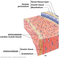
Pericardial Cavity and Pericardium
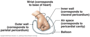
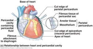
The heart is enclosed within the pericardial sac, which protects and anchors the heart while allowing movement during contraction.
Fibrous pericardium: Outermost protective layer
Parietal pericardium: Lines the inner surface of the pericardial sac
Visceral pericardium (Epicardium): Covers the heart's surface
Pericardial cavity: Space between parietal and visceral layers, containing pericardial fluid to reduce friction
Histology of the Heart Wall
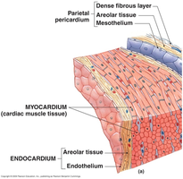
Each layer of the heart wall has a specific structure and function:
Epicardium: Produces serous fluid, protects the heart's exterior
Myocardium: Thickest layer, responsible for contraction
Endocardium: Smooth lining for chambers, continuous with blood vessel endothelium
Gross Anatomy of the Heart
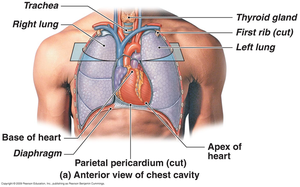
External Features and Landmarks
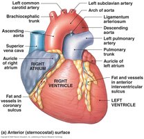
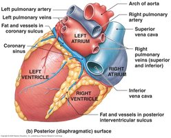
The heart has four chambers and several external landmarks that are important for orientation and function.
Right atrium: Receives blood from systemic circuit
Right ventricle: Pumps blood to pulmonary circuit
Left atrium: Receives blood from pulmonary circuit
Left ventricle: Pumps blood to systemic circuit
Coronary sulcus: Groove separating atria and ventricles
Anterior and posterior interventricular sulci: Boundaries between ventricles
Auricles: Flaps on atria
Major vessels: Superior/inferior vena cava, aorta, pulmonary trunk, pulmonary arteries and veins
Ligamentum arteriosum: Remnant of fetal ductus arteriosus
Internal Features and Landmarks
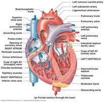
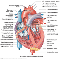
The internal anatomy of the heart includes septa, valves, and specialized muscle structures that ensure unidirectional blood flow and efficient contraction.
Interatrial septum: Separates right and left atria
Interventricular septum: Separates right and left ventricles
Valves: Tricuspid (right AV), bicuspid/mitral (left AV), pulmonary, and aortic valves
Chordae tendineae: Connect valve cusps to papillary muscles
Trabeculae carneae: Muscular ridges in ventricles
Moderator band: Conducts electrical signals in right ventricle
Right Atrium
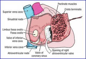
The right atrium receives deoxygenated blood from the body and heart wall. It contains several important anatomical features:
Openings: Superior vena cava, inferior vena cava, coronary sinus
Foramen ovale (fetus): Connects atria during fetal development; becomes fossa ovalis after birth
Pectinate muscles: Muscular ridges in anterior wall
Auricle: Flap that increases atrial volume
Right Ventricle
The right ventricle pumps blood into the pulmonary circuit. Key features include:
Trabeculae carneae: Muscular ridges
Moderator band: Conducts electrical impulses
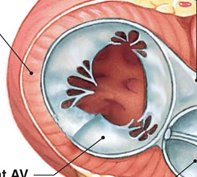
Tricuspid valve: Three cusps, prevents backflow into atrium
Chordae tendineae and papillary muscles: Prevent valve prolapse during contraction
Right Ventricle Outflow
The conus arteriosus leads to the pulmonary valve and trunk, directing blood to the lungs.
Pulmonary valve: Prevents backflow into ventricle
Pulmonary trunk: Splits into left and right pulmonary arteries
Left Atrium and Left Ventricle
The left atrium receives oxygenated blood from the lungs, while the left ventricle pumps it into the systemic circuit. The left ventricle has a thicker wall to generate higher pressure.
Left atrium: Receives blood from pulmonary veins, contains auricle
Bicuspid (mitral) valve: Two cusps, prevents backflow into atrium
Left ventricle: Thicker myocardium, no moderator band, pumps blood through aortic valve into aorta
Ligamentum arteriosum: Connects pulmonary trunk to aorta (fetal remnant)
Aortic Sinuses
Located at the base of the ascending aorta, these sacs prevent valve cusps from sticking to the aorta and are the origin of the coronary arteries.
Heart Valves and Cardiac Skeleton
Atrioventricular (AV) and Semilunar Valves
Valves ensure unidirectional blood flow through the heart and prevent backflow.
AV valves: Between atria and ventricles; open during ventricular relaxation
Semilunar valves: Pulmonary and aortic; open during ventricular contraction
Chordae tendineae and papillary muscles: Prevent AV valve prolapse
Valve disease: Inflammation (carditis) can lead to valvular heart disease (VHD)
Cardiac Skeleton
The cardiac skeleton is a framework of connective tissue that supports the heart's valves and electrically insulates the atria from the ventricles.
Provides structural support and elasticity
Stabilizes valves and prevents overexpansion
Electrically insulates atrial and ventricular muscle fibers
Coronary Circulation
Coronary Arteries
The coronary arteries supply oxygenated blood to the heart muscle itself. They originate from the aortic sinuses.
Right coronary artery: Supplies right atrium, portions of both ventricles, and conduction system
Left coronary artery: Supplies left ventricle, left atrium, and interventricular septum
Main branches: Circumflex artery, anterior interventricular artery, marginal arteries, posterior interventricular artery
Coronary Veins
Coronary veins collect deoxygenated blood from the myocardium and return it to the right atrium via the coronary sinus.
Great cardiac vein: Runs in anterior interventricular sulcus
Coronary sinus: Main venous drainage into right atrium
Other veins: Posterior cardiac, middle cardiac, small cardiac, anterior cardiac veins
Summary Table: Heart Wall Layers
Layer | Tissue Type | Function |
|---|---|---|
Endocardium | Simple squamous epithelium, areolar tissue | Lines chambers, protects myocardium |
Myocardium | Cardiac muscle tissue | Contraction, pumping action |
Epicardium (Visceral Pericardium) | Mesothelium, areolar tissue | Protects, produces serous fluid |
Parietal Pericardium | Mesothelium, areolar, dense fibrous tissue | Protects, anchors heart |
Review Checklist
Cardiac muscle cell structure and function
Heart wall layers and tissue types
External and internal heart features
Heart valves and cardiac skeleton
Coronary circulation: arteries and veins
