 뒤로
뒤로Anatomy of the Heart: Structure, Circuits, and Internal Features (A&P II: Lab 3 Review)
스터디 가이드 - 스마트 노트
 자료에 맞춘 맞춤형 노트, 핵심 정의, 예시, 맥락을 확장해 제공합니다.
The Heart: Structure and Circulation
Overview of the Heart
The heart is a muscular organ responsible for pumping blood throughout the body via two main circuits: the pulmonary and systemic circuits. It is composed of four chambers and is surrounded by protective layers collectively known as the pericardium.
The Pericardium
Layers of the Pericardium
The pericardium is a double-walled sac that encloses the heart, providing protection and reducing friction as the heart beats. The two main layers are:
Parietal Pericardium: The outer fibrous layer that anchors the heart in the thoracic cavity.
Epicardium (Visceral Pericardium): The inner serous layer that lies directly on the heart surface.
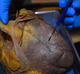
External Features of the Heart
Major Chambers and Sulci
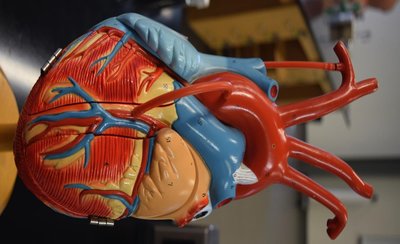
The heart's external anatomy includes four chambers (right and left atria, right and left ventricles) and visible grooves called sulci, which mark the boundaries between chambers and house coronary vessels.
Anterior Interventricular Sulcus: Marks the boundary between the right and left ventricles on the anterior surface.
Posterior Interventricular Sulcus: Marks the boundary on the posterior surface.
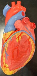
Pulmonary and Systemic Circuits
Pulmonary Circuit
The pulmonary circuit carries deoxygenated blood from the right side of the heart to the lungs and returns oxygenated blood to the left side of the heart.
Right Atrium: Receives deoxygenated blood from the superior and inferior vena cava.
Tricuspid Valve: Prevents backflow into the right atrium.
Right Ventricle: Pumps blood through the pulmonary semilunar valve into the pulmonary trunk and arteries to the lungs.
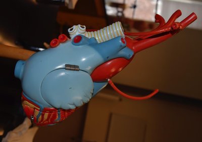
Systemic Circuit
The systemic circuit delivers oxygenated blood from the left side of the heart to the body and returns deoxygenated blood to the right side.
Left Atrium: Receives oxygenated blood from the pulmonary veins.
Bicuspid (Mitral) Valve: Prevents backflow into the left atrium.
Left Ventricle: Pumps blood through the aortic semilunar valve into the aorta and systemic arteries.
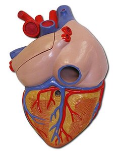
Major Blood Vessels Associated with the Heart
Arteries and Veins
The heart is connected to major blood vessels that facilitate blood flow to and from the body and lungs:
Aorta: The main artery carrying oxygenated blood to the systemic circuit.
Pulmonary Trunk and Arteries: Carry deoxygenated blood to the lungs.
Pulmonary Veins: Return oxygenated blood from the lungs to the left atrium.
Vena Cavae: Superior and inferior veins returning deoxygenated blood to the right atrium.
Internal Structures of the Heart
Heart Wall Layers
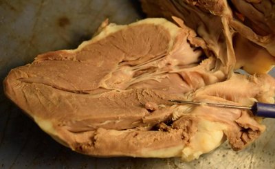
The heart wall consists of three layers:
Endocardium: The innermost layer lining the heart chambers and valves.
Myocardium: The thick, muscular middle layer responsible for contraction.
Epicardium: The outermost layer, also known as the visceral pericardium.
Heart Valves
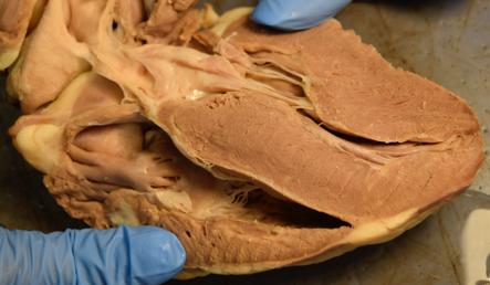
Valves ensure unidirectional blood flow through the heart:
Atrioventricular (AV) Valves: Tricuspid (right) and bicuspid/mitral (left) valves between atria and ventricles.
Semilunar Valves: Pulmonary (right) and aortic (left) valves at the exits of the ventricles.
Chordae Tendineae and Papillary Muscles
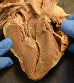
These structures anchor the AV valves and prevent prolapse during ventricular contraction:
Chordae Tendineae: Tendinous cords connecting valve leaflets to papillary muscles.
Papillary Muscles: Muscular projections from the ventricular wall.
Coronary Circulation
Supplying the Heart Muscle
The heart muscle (myocardium) receives oxygen and nutrients from the coronary arteries, which branch off the aorta. Deoxygenated blood is collected by coronary veins and returned to the right atrium via the coronary sinus.
Coronary Arteries: Supply oxygen-rich blood to the myocardium.
Coronary Sinus: Collects deoxygenated blood from the myocardium.
Summary Table: Heart Chambers, Valves, and Major Vessels
Chamber | Valve | Major Vessel In | Major Vessel Out |
|---|---|---|---|
Right Atrium | Tricuspid Valve | Superior/Inferior Vena Cava | Right Ventricle |
Right Ventricle | Pulmonary Semilunar Valve | Right Atrium | Pulmonary Trunk/Arteries |
Left Atrium | Bicuspid (Mitral) Valve | Pulmonary Veins | Left Ventricle |
Left Ventricle | Aortic Semilunar Valve | Left Atrium | Aorta |
Key Equations
Cardiac Output (CO): The volume of blood pumped by each ventricle per minute.
Where HR is heart rate (beats per minute) and SV is stroke volume (mL per beat).
Additional info: The ligamentum arteriosum is a remnant of the fetal ductus arteriosus, which connects the pulmonary trunk to the aorta during fetal development. After birth, it closes and becomes a ligament.
