 뒤로
뒤로BIO 201: The Human Body – Foundations of Anatomy & Physiology
스터디 가이드 - 스마트 노트
 자료에 맞춘 맞춤형 노트, 핵심 정의, 예시, 맥락을 확장해 제공합니다.
The Human Body: An Orientation
Introduction to Anatomy & Physiology
Anatomy and Physiology (A&P) is the foundational study of the human body’s structure (anatomy) and function (physiology). Understanding both disciplines is essential for health sciences, as form and function are deeply interconnected.
Anatomy: The study of the structure and form of the body and its parts.
Physiology: The study of how the body and its parts function.
Form and Function: Anatomical structures are designed to perform specific functions; understanding one requires knowledge of the other.
Subdivisions of Anatomy
Microscopic Anatomy: Structures too small to be seen with the naked eye.
Cytology: Study of cells and their internal structure.
Histology: Study of tissues.
Gross (Macroscopic) Anatomy: Structures visible to the naked eye.
Systemic Anatomy: Study of body systems.
Regional Anatomy: Study of specific regions of the body.
Surface Anatomy: Study of external features and their relation to deeper structures.
Comparative Anatomy: Comparison of structures across species.
Embryology: Study of development from conception to birth.
Pathologic Anatomy: Changes due to disease.
Radiographic Anatomy: Structures visualized by imaging techniques.
Subdivisions of Physiology
Cardiovascular Physiology: Functioning of the heart and blood vessels.
Neurophysiology: Functioning of nerves and nervous system organs.
Respiratory Physiology: Functioning of respiratory organs.
Reproductive Physiology: Functioning of reproductive hormones and cycles.
Pathophysiology: Function during disease or injury.
Levels of Biological Organization
Biological Hierarchy
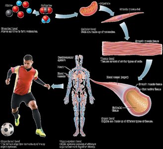
The human body is organized into a hierarchy of increasing complexity:
Chemical Level: Atoms and molecules.
Cellular Level: Cells, the basic units of life.
Tissue Level: Groups of similar cells performing common functions.
Organ Level: Two or more tissue types working together.
Organ System Level: Related organs working together.
Organismal Level: All organ systems functioning together in the human body.
Surface Area to Volume Ratio
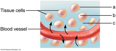
As organisms increase in size, their surface area to volume ratio decreases, affecting exchange of materials and heat regulation.
Small cells have a high surface area to volume ratio, facilitating efficient exchange with the environment.
Larger organisms require specialized systems for exchange and transport.

Necessary Life Functions
Characteristics of Living Things
Organization: Complex structure and order.
Metabolism: All chemical reactions in the body.
Anabolism: Building larger molecules from smaller ones.
Catabolism: Breaking down large molecules into smaller ones.
Growth and Development: Assimilation of materials and increase in size or specialization.
Responsiveness: Ability to sense and react to stimuli.
Regulation: Adjusting internal functions to environmental changes (homeostasis).
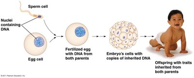
Reproduction: Producing new cells for growth, maintenance, repair, and offspring.
Examples of Necessary Life Functions
Maintaining Boundaries: Separation between internal and external environments (e.g., plasma membrane, skin).
Digestion and Absorption: Breaking down food and absorbing nutrients.
Metabolism: All chemical reactions, including energy production and use.
Excretion: Removal of waste products.
Reproduction: Cellular and organismal reproduction.
Growth: Increase in size and complexity.
Responsiveness and Movement: Reacting to stimuli and moving body parts.
Homeostasis
Definition and Importance
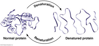
Homeostasis is the process by which the body maintains a stable internal environment despite changes in external conditions. It is essential for the survival and optimal functioning of cells and proteins.
Maintaining homeostasis ensures that temperature, pH, and other conditions remain within narrow limits.
Proteins require specific conditions to maintain their shape and function; deviations can lead to denaturation and loss of function.
Thermoregulation Example
Body temperature is regulated around a set point (about 98.6°F or 37°C).
Responses to heat: sweating, vasodilation, seeking shade.
Responses to cold: shivering, vasoconstriction, seeking warmth.


Feedback Mechanisms
Negative Feedback: Most common; a change in a variable triggers a response that counteracts the initial change, maintaining homeostasis.
Examples: regulation of body temperature, blood glucose, blood pressure.
Positive Feedback: Amplifies the initial stimulus; less common and usually associated with specific events (e.g., childbirth, blood clotting).
Not paired with antagonistic mechanisms and does not maintain a set point.
Components of Homeostatic Control
Receptor: Detects changes in the environment (stimuli).
Control Center: Processes information and determines the response.
Effector: Carries out the response to restore homeostasis.
Organ Systems Overview
The 11 Organ Systems of the Human Body
Integumentary System
Skeletal System
Muscular System
Nervous System
Endocrine System
Cardiovascular System
Lymphatic System
Respiratory System
Urinary System
Digestive System
Male and Female Reproductive Systems
Anatomic Position and Directional Terms
Anatomic Position
The standard reference position for the body in the study of anatomy:
Upright stance, feet parallel and flat, arms at sides, palms facing forward, head level, eyes forward.
Anatomic Planes and Sections
Coronal (Frontal) Plane: Divides body into anterior (front) and posterior (back) parts.
Transverse (Horizontal) Plane: Divides body into superior (top) and inferior (bottom) parts.
Sagittal Plane: Divides body into left and right parts (midsagittal = equal halves).
Directional Terms
Term | Meaning | Example |
|---|---|---|
Anterior | In front of; toward the front | The stomach is anterior to the spinal cord |
Posterior | In back of; toward the back | The heart is posterior to the sternum |
Superior | Closer to the head | The chest is superior to the pelvis |
Inferior | Closer to the feet | The stomach is inferior to the heart |
Medial | Toward the midline | The lungs are medial to the shoulders |
Lateral | Away from the midline | The arms are lateral to the heart |
Proximal | Closer to point of attachment | The elbow is proximal to the hand |
Distal | Farther from point of attachment | The wrist is distal to the elbow |
Superficial | Closer to the surface | The skin is superficial to muscles |
Deep | Farther from the surface | The heart is deep to the rib cage |
Body Cavities and Membranes
Major Body Cavities
Posterior Aspect: Cranial cavity (houses brain), vertebral canal (houses spinal cord); encased in bone.
Ventral Cavity: Thoracic cavity (superior), abdominopelvic cavity (inferior); not completely encased in bone.
Serous Membranes
Line the ventral cavity subdivisions.
Two layers:
Parietal Layer: Lines internal surface of body wall.
Visceral Layer: Covers external surface of organs.
Serous Cavity: Space between layers, filled with serous fluid to reduce friction.
Examples of Serous Membranes
Pericardium: Surrounds the heart.
Pleura: Surrounds the lungs.
Peritoneum: Lines the abdominopelvic cavity and covers abdominal organs.
Medical Imaging Techniques
Overview of Imaging Methods
Technique | Description | Advantages | Disadvantages |
|---|---|---|---|
Radiography (X-ray) | High-energy radiation passes through body | Good for bones, teeth, tumors | Radiation risk, overlapping organs |
Ultrasound | High-frequency sound waves reflect off organs | No radiation, portable, inexpensive | Less sharp images |
Digital Subtraction Angiography (DSA) | 3D X-ray with contrast for blood vessels | Detects blockages, useful for angioplasty | Requires injection, not fully noninvasive |
Computed Tomography (CT) | Low-intensity X-rays, computer-generated slices | Sharp, 3D images, little overlap | Higher radiation dose |
Magnetic Resonance Imaging (MRI) | Magnetic fields and radio waves | Excellent for soft tissues | Claustrophobia, dense tissues less visible |
Positron Emission Tomography (PET) | Radioactive glucose, detects metabolic activity | Assesses tissue activity, cancer detection | Radioactive tracer required |
Summary Table: Levels of Organization
Level | Description | Example |
|---|---|---|
Chemical | Atoms and molecules | Water, proteins |
Cellular | Basic unit of life | Muscle cell |
Tissue | Group of similar cells | Muscle tissue |
Organ | Two or more tissues | Heart |
Organ System | Related organs | Cardiovascular system |
Organismal | All organ systems | Human body |
Additional info: This guide covers the foundational concepts of human anatomy and physiology, including organization, necessary life functions, homeostasis, organ systems, anatomical terminology, body cavities, and medical imaging. These topics align with Chapters 1–4 of a standard Anatomy & Physiology curriculum.
