 뒤로
뒤로Blood Groups, Leukocytes, and Hemostasis: Study Notes for Anatomy & Physiology
스터디 가이드 - 스마트 노트
 자료에 맞춘 맞춤형 노트, 핵심 정의, 예시, 맥락을 확장해 제공합니다.
Blood Groups & Typing
Antigens and Antibodies
Blood group antigens and antibodies are essential for distinguishing self from non-self and play a critical role in transfusion medicine and immune responses.
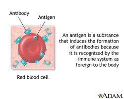
Antigens (agglutinogens): Unique molecules on the surface of red blood cells (RBCs) that are recognized by the immune system. Foreign antigens can trigger an immune response.
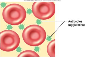
Antibodies (agglutinins): Proteins secreted by immune cells in response to foreign antigens. They bind specifically to antigens and can cause agglutination (clumping) of RBCs.
Agglutination: The process where antibodies bind to antigens on RBCs, causing them to clump together. This is the basis for blood typing and transfusion reactions.
Blood Group Antigens
The specific antigens present on the surface of RBCs determine an individual's blood type. The most clinically significant blood group systems are the ABO and Rh systems.
Type O: Most common blood type; lacks both A and B antigens.
Type AB: Rarest blood type; has both A and B antigens.
ABO Blood Typing
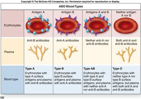
The ABO system classifies blood based on the presence or absence of A and B antigens on RBCs and corresponding antibodies in plasma.
Blood Type | Antigens on RBCs | Antibodies in Plasma |
|---|---|---|
Type A | A | Anti-B |
Type B | B | Anti-A |
Type AB | A and B | None |
Type O | None | Anti-A and Anti-B |
Blood Typing Procedure
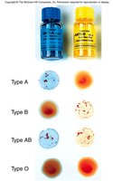
Blood typing involves mixing a blood sample with anti-A and anti-B sera to observe agglutination, which indicates the presence of specific antigens.
Transfusion Reaction
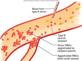
If incompatible blood is transfused, antibodies in the recipient's plasma bind to donor RBC antigens, causing agglutination and hemolysis. This can block blood vessels and lead to kidney failure or death.
Clinical Case Example
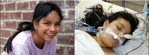
Errors in blood typing or organ matching can have fatal consequences, as illustrated by the tragic case of Jesica Santillan, who received mismatched organs (Type O recipient, Type A donor).

Other Blood Groups
There are over 100 other blood group antigens (e.g., MN, Duffy, Kell, Kidd, Lewis), but these rarely cause transfusion reactions compared to ABO and Rh systems.
Rh Group
The Rh (D) antigen is another major blood group antigen. Individuals with the antigen are Rh positive (Rh+); those without are Rh negative (Rh-). Rh antibodies are not naturally present but can form after exposure to Rh+ blood.
85% of white Americans are Rh+; 99% of Asians are Rh+.
Rh- individuals can develop anti-D antibodies after exposure (e.g., transfusion or pregnancy).
Hemolytic Disease of the Newborn (HDN)
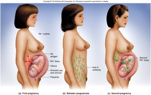
HDN occurs when an Rh- mother develops anti-D antibodies after exposure to Rh+ fetal blood, which can attack the RBCs of a subsequent Rh+ fetus, causing anemia and neurological damage. Prevention involves administering RhoGAM to Rh- mothers.
Universal Donors and Recipients
Universal donor: Type O- (lacks A, B, and Rh antigens).
Universal recipient: Type AB+ (lacks anti-A, anti-B, and anti-Rh antibodies).
Leukocytes (White Blood Cells, WBCs)
General Features
Leukocytes are immune cells that protect the body against pathogens. They have a conspicuous nucleus, mature in lymphoid organs, and migrate from blood to connective tissues. Normal count: 5,000–10,000/μL.
Classification of Leukocytes
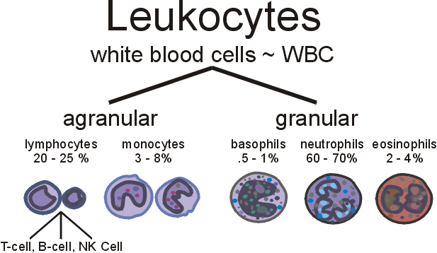
Leukocytes are classified as granulocytes or agranulocytes based on the presence of cytoplasmic granules.
Type | Examples | Percentage of WBCs |
|---|---|---|
Agranulocytes | Lymphocytes, Monocytes | 20–30%, 3–8% |
Granulocytes | Neutrophils, Eosinophils, Basophils | 60–70%, 2–4%, 0.5–1% |
Agranulocytes
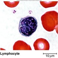
Lymphocytes: Round, dark blue nucleus; produce antibodies; include B cells, T cells, and natural killer cells.
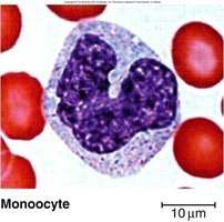
Monocytes: Large, kidney-shaped nucleus; differentiate into macrophages; increase in viral infections and inflammation.
Granulocytes
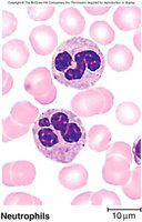
Neutrophils: Pale lilac granules, 3–5 lobed nucleus; increase during bacterial infections.
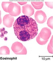
Eosinophils: Red-orange granules, bilobed nucleus; increase with allergies and parasitic diseases.
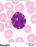
Basophils: Large, dark violet granules, S-shaped nucleus; granules contain heparin and histamine.
Leukopoiesis and Leukocyte Disorders
Leukopoiesis
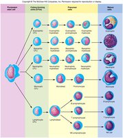
Leukopoiesis is the process of white blood cell formation from hematopoietic stem cells in the bone marrow.
Leukocyte Disorders
Leukopenia: Low WBC count (<5,000/μL); increases risk of infection.
Leukocytosis: High WBC count (>10,000/μL); often due to infection, allergy, or disease.
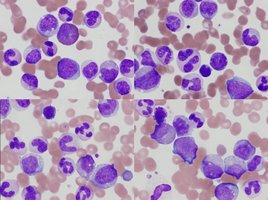
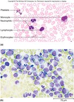
Leukemia: Cancer of hematopoietic tissue; uncontrolled WBC production disrupts normal cell percentages and impairs clotting.
Platelets and Hemostasis
Platelets
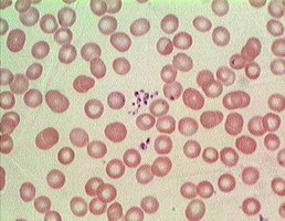
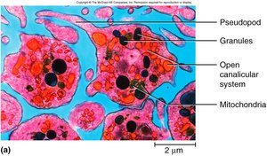
Platelets are small cell fragments (2–4 μm) derived from megakaryocytes. They play a vital role in hemostasis and have a lifespan of 5–9 days. Normal count: 130,000–400,000/μL.
Platelet Functions
Plug small injuries to blood vessels
Secrete clotting factors
Release serotonin (vasoconstriction)
Dissolve old clots
Attract WBCs
Phagocytize bacteria
Thrombopoiesis (Platelet Production)
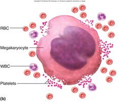
Platelets are produced from megakaryocytes in the bone marrow. Stem cells differentiate into megakaryoblasts, which become megakaryocytes. Cytoplasmic fragments of megakaryocytes enter the bloodstream as platelets.
Hemostasis
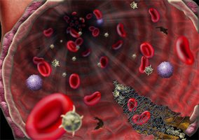
Hemostasis is the process of stopping bleeding. It involves three main steps:
Vascular spasm: Vasoconstriction reduces blood flow.
Platelet plug formation: Platelets adhere to exposed collagen and aggregate.
Coagulation: Fibrin forms a mesh that traps blood cells, forming a stable clot.
Coagulation Pathways
Blood coagulation occurs via two pathways:
Extrinsic pathway: Triggered by external trauma; rapid (12–15 seconds).
Intrinsic pathway: Triggered by internal vessel damage; slower (30–45 seconds).
Both pathways require calcium ions (Ca2+).
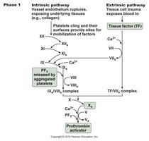
Completion of Coagulation
The final steps of coagulation involve the conversion of prothrombin to thrombin, which then converts fibrinogen to fibrin, forming the clot. Thrombin also accelerates its own formation (positive feedback).
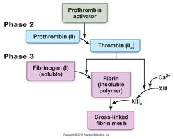
Fate of Blood Clots
After clot formation, clot retraction occurs within 30 minutes. Fibrinolysis (clot dissolution) is mediated by plasmin, a fibrin-dissolving enzyme. This process is also regulated by positive feedback.
Prevention of Inappropriate Clotting
Platelet repulsion: Platelets do not adhere to undamaged endothelium.
Thrombin dilution: Rapid blood flow prevents clot formation; slow flow (e.g., in shock) increases risk.
Natural anticoagulants: Heparin inhibits thrombin formation.
Coagulation Disorders
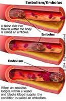
Embolus: A clot traveling in a vessel.
Thrombosis: Abnormal clotting in unbroken vessels, often in leg veins of inactive people.
Pulmonary embolism: A clot that travels to the lungs.
Infarction: Tissue death due to blocked blood supply (e.g., myocardial infarction, stroke).
Prevention: Aspirin, heparin, and warfarin (Coumadin) are used to reduce clot risk.
Disseminated Intravascular Coagulation (DIC)
DIC is a pathological process involving widespread clotting and bleeding due to consumption of clotting factors and platelets. Causes include pregnancy, cancers, and septicemia.
Hemophilia
Hemophilia is a genetic disorder (often X-linked recessive) characterized by a deficiency of clotting factors, leading to excessive bleeding and pain. Treatment involves transfusion of plasma or purified clotting factors.
