 뒤로
뒤로Blood: Leukocytes, Hemostasis, and Blood Disorders
스터디 가이드 - 스마트 노트
 자료에 맞춘 맞춤형 노트, 핵심 정의, 예시, 맥락을 확장해 제공합니다.
Leukocytes: Production, Function, and Disorders
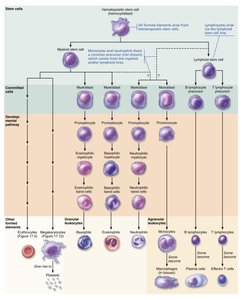
Leukopoiesis: Production and Life Span of Leukocytes
Leukopoiesis is the process of white blood cell (WBC) production, essential for immune defense. Leukocytes are divided into granulocytes and agranulocytes, each with distinct developmental pathways and life spans.
Granulocyte Production: Originates from myeloid stem cells and progresses through several stages: myeloblast, promyelocyte, myelocyte, band cell, and mature granulocyte. Granulocytes are stored in bone marrow and have a short life span (hours to days).
Agranulocyte Production: Monocytes and neutrophils share a precursor, while lymphocytes derive from the lymphoid line. Monocytes can live for months, and lymphocytes can persist for hours to decades.
Regulation: Interleukins (e.g., IL-3, IL-5) and colony-stimulating factors (CSFs) regulate leukopoiesis, stimulating specific WBC lineages.
Monocytes and Lymphocytes: Interaction and Immune Function
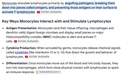
Monocytes stimulate lymphocytes by engulfing pathogens, breaking them into antigens, and presenting these antigens to activate T-lymphocytes. This process is crucial for adaptive immunity.
Antigen Presentation: Monocytes (and macrophages) display antigens on MHC molecules to activate T-cells.
Cytokine Production: Monocytes release cytokines (e.g., interleukins) that direct lymphocyte behavior.
Differentiation: Monocytes become macrophages in tissues, interacting closely with lymphocytes to coordinate immune responses.
Leukocyte Disorders
Disorders of leukocytes can involve overproduction, underproduction, or abnormal function, impacting immune defense and overall health.
Leukemias: Cancers of WBCs, classified by rate (acute or chronic) and cell type (myeloid or lymphocytic). Acute forms progress rapidly and affect children; chronic forms are slower and more common in older adults. Untreated, leukemias are fatal due to nonfunctional WBCs crowding out other blood cells.
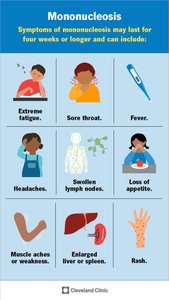
Infectious Mononucleosis: A viral disease (Epstein-Barr virus) causing excess, enlarged lymphocytes. Symptoms include fatigue, sore throat, and fever, typically resolving in 4–6 weeks.
Leukopenia: Abnormally low WBC count, often drug-induced, leading to increased infection risk.
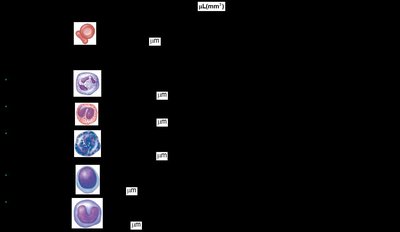
Summary Table: Formed Elements of Blood
The following tables summarize the identification, functions, and normal counts of the formed elements of blood.
Cell Type | Identification | Function | Cells per µL |
|---|---|---|---|
Erythrocytes | Biconcave, anucleate | Transport O2 and CO2 | 4–6 million |
Neutrophils | Multilobed nucleus, pale granules | Phagocytize bacteria | 3,000–7,000 |
Eosinophils | Bilobed nucleus, red granules | Kill parasitic worms; allergy modulation | 100–400 |
Basophils | Lobed nucleus, large blue granules | Release histamine; inflammation | 20–50 |
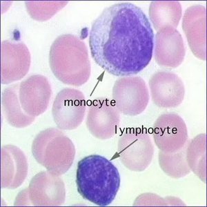
Lymphocytes | Large nucleus, thin rim of cytoplasm | Immune response via T and B cells | 1,500–3,000 |
Monocytes | Kidney-shaped nucleus | Phagocytosis; develop into macrophages | 100–700 |
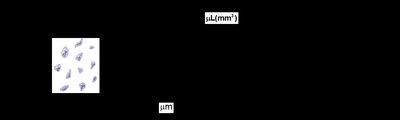
Hemostasis: Mechanisms to Prevent Blood Loss
Overview of Hemostasis
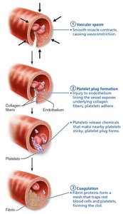
Hemostasis is the process that stops bleeding through a rapid series of reactions. It involves three major steps: vascular spasm, platelet plug formation, and coagulation.
Step 1: Vascular Spasm – Vasoconstriction of the injured vessel reduces blood loss, triggered by injury, chemicals, and pain reflexes.
Step 2: Platelet Plug Formation – Platelets adhere to exposed collagen, become activated, and aggregate to form a temporary plug. von Willebrand factor aids adhesion; ADP, serotonin, and thromboxane A2 amplify aggregation.
Step 3: Coagulation – Fibrin reinforces the platelet plug, forming a stable blood clot through a cascade of clotting factors.
Coagulation Pathways
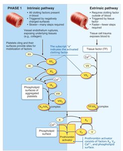
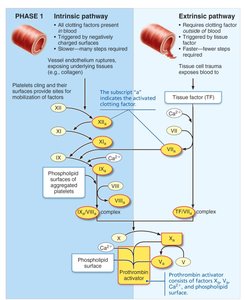
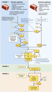
Coagulation involves a cascade of plasma proteins (clotting factors) that ultimately convert fibrinogen to fibrin. There are two initial pathways—intrinsic and extrinsic—that converge at the common pathway.
Intrinsic Pathway: Initiated by factors within the blood (e.g., activated platelets, collagen). Slower, involves more steps.
Extrinsic Pathway: Triggered by tissue factor (TF) from damaged tissues. Faster, fewer steps.
Both pathways activate Factor X, which, with Ca2+, PF3, and Factor V, forms prothrombin activator.
Common Pathway and Clot Formation
Once prothrombin activator is formed, the cascade proceeds through two additional phases:
Phase 2: Prothrombin is converted to thrombin.
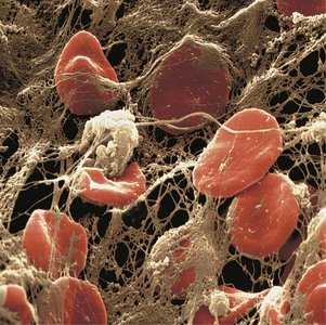
Phase 3: Thrombin converts soluble fibrinogen into insoluble fibrin, forming a mesh that stabilizes the clot. Factor XIII cross-links fibrin, strengthening the clot.

Blood Clotting Factors (Procoagulants)
Clotting factors are mostly plasma proteins produced by the liver and are essential for the coagulation cascade. Some require vitamin K for synthesis.
Factor Number | Name | Nature | Source | Function |
|---|---|---|---|---|
I | Fibrinogen | Plasma protein | Liver | Converted to fibrin |
II | Prothrombin | Plasma protein | Liver* | Converted to thrombin |
III | Tissue factor (TF) | Glycoprotein | Tissue cells | Activates extrinsic pathway |
IV | Calcium ions | Inorganic ion | Plasma | Needed for all stages |
V | Proaccelerin | Plasma protein | Liver, platelets | Common pathway |
VII | Proconvertin | Plasma protein | Liver* | Extrinsic/intrinsic pathways |
VIII | Antihemophilic factor | Plasma protein | Liver, lung capillaries | Intrinsic pathway |
IX | Plasma thromboplastin component | Plasma protein | Liver* | Intrinsic pathway |
X | Stuart factor | Plasma protein | Liver* | Common pathway |
XI | Plasma thromboplastin antecedent | Plasma protein | Liver | Intrinsic pathway |
XII | Hageman factor | Plasma protein | Liver | Intrinsic pathway, inflammation |
XIII | Fibrin stabilizing factor | Plasma protein | Liver, bone marrow | Cross-links fibrin |
Additional info: Factors II, VII, IX, and X require vitamin K for synthesis.
Regulation and Limitation of Clot Formation
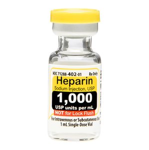
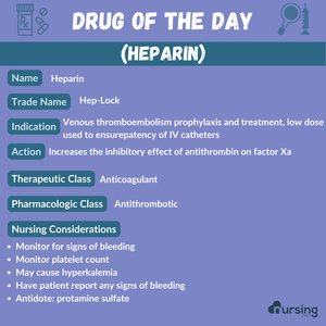
Clot growth is limited by swift removal and inhibition of clotting factors. Antithrombin III and heparin (from basophils and mast cells) inactivate thrombin and other factors, preventing excessive clotting. Endothelial cells secrete antithrombotic substances (nitric oxide, prostacyclin) to prevent platelet adhesion to intact vessels.
Disorders of Hemostasis
Thromboembolic Disorders
These disorders involve undesirable clot formation:
Thrombus: Clot in an unbroken vessel, may block circulation and cause tissue death.
Embolus: Freely floating clot; can lodge in vessels (embolism), causing pulmonary or cerebral complications.
Anticoagulant Drugs: Aspirin, heparin, and warfarin are used to prevent or treat thromboembolic conditions.
Bleeding Disorders
Bleeding disorders result from deficiencies in platelets or clotting factors:
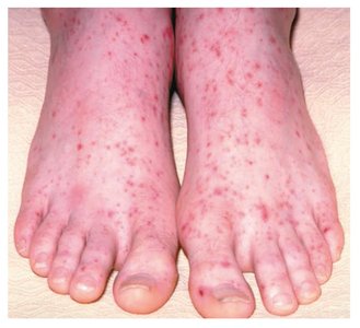
Thrombocytopenia: Low platelet count, leading to petechiae (small hemorrhages) and increased bleeding risk. Causes include bone marrow suppression or destruction.
Impaired Liver Function: Reduces synthesis of clotting factors, often due to vitamin K deficiency, hepatitis, or cirrhosis.
Hemophilia: Hereditary bleeding disorders due to deficiencies in factors VIII (A), IX (B), or XI (C). Treated with recombinant clotting factors.
Disseminated Intravascular Coagulation (DIC)
DIC involves both widespread clotting and severe bleeding, often triggered by sepsis, incompatible transfusions, or pregnancy complications. Clotting factors are depleted, leading to uncontrolled bleeding.
Blood Typing and Transfusion
ABO and Rh Blood Groups
Blood type is determined by antigens (agglutinogens) on erythrocyte surfaces. The ABO system is based on the presence of A and/or B antigens; the Rh system is based on the D antigen.
Antibodies: Plasma contains antibodies against absent antigens (e.g., type A blood has anti-B antibodies).
Transfusion Compatibility: Universal donor: O-; universal recipient: AB+.
Transfusion Reactions: Occur if incompatible blood is transfused, causing hemolysis and possible kidney failure.
Hemolytic Disease of the Newborn: Occurs when an Rh-negative mother carries an Rh-positive fetus; prevented by RHOGAM administration.
Blood Loss and Replacement
The cardiovascular system compensates for blood loss by vasoconstriction and increased RBC production. Severe loss (>30%) can cause shock. Volume is restored with saline or electrolyte solutions, but oxygen-carrying capacity requires RBC transfusion.
Blood Tests and Clinical Insights
Blood tests provide diagnostic information:
Hematocrit: Low values indicate anemia.
Blood Glucose: Screens for diabetes.
Differential WBC Count: Identifies infections or immune disorders.
Prothrombin Time (PT) and Platelet Count: Assess hemostasis.
Comprehensive Metabolic Panel (CMP): Evaluates organ function.
Complete Blood Count (CBC): Measures all formed elements and hemoglobin.
Developmental Aspects of Blood
Fetal blood cells originate in the yolk sac, liver, and spleen before red bone marrow becomes the primary site. Fetal hemoglobin (hemoglobin F) has a higher affinity for oxygen than adult hemoglobin. Blood disorders increase with age, often due to cardiovascular or immune system diseases.
