 뒤로
뒤로Blood: Structure, Function, and Clinical Relevance – Study Notes for Anatomy & Physiology
스터디 가이드 - 스마트 노트
 자료에 맞춘 맞춤형 노트, 핵심 정의, 예시, 맥락을 확장해 제공합니다.
Blood and the Cardiovascular System
Overview of the Cardiovascular System
The cardiovascular system is essential for transporting substances throughout the body. It consists of three main components:
Blood – the fluid connective tissue that transports materials.
Heart – the muscular pump that circulates blood.
Blood vessels – the network of conduits that carry blood to and from tissues.
Components and Functions of Blood
Main Components of Blood
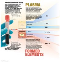
Blood is a specialized connective tissue composed of cells suspended in a fluid matrix. It consists of:
Plasma – the liquid matrix, making up 46–63% of blood volume.
Formed elements – cells and cell fragments, including red blood cells (RBCs), white blood cells (WBCs), and platelets.
Functions of Blood
Transport of gases (O2, CO2), nutrients, hormones, and metabolic wastes.
Regulation of pH and ion composition of interstitial fluids.
Restriction of fluid losses at injury sites (hemostasis).
Defense against toxins and pathogens (immune response).
Stabilization of body temperature by redistributing heat.
Physical Characteristics of Blood
Temperature: Slightly higher than body temperature.
Viscosity: Higher than water due to cells and proteins.
pH: Slightly alkaline (7.35–7.45).
Volume: About 7% of body weight in liters.
Fractionation is the process of separating blood into plasma and formed elements for analysis.
Plasma Composition
92% water
7% plasma proteins (albumins, globulins, fibrinogen)
1% other solutes (nutrients, wastes, electrolytes)
Plasma is similar to interstitial fluid but has a higher concentration of dissolved proteins and different gas concentrations.
Plasma Proteins
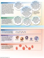
Albumins: Most abundant; maintain osmotic pressure and transport substances.
Globulins: Include antibodies (immunoglobulins) and transport proteins.
Fibrinogen: Soluble protein involved in clotting; converted to fibrin during coagulation.
Most plasma proteins are synthesized by the liver, except immunoglobulins, which are produced by WBCs.
Formed Elements
Red blood cells (erythrocytes)
White blood cells (leukocytes)
Platelets (thrombocytes)
Hemopoiesis is the process of producing formed elements, primarily in red bone marrow.
Red Blood Cells (Erythrocytes)
Characteristics and Functions
Make up 99.9% of formed elements.
Contain hemoglobin, a red pigment that binds and transports oxygen and carbon dioxide.
Normal RBC count: Adult males 4.5–6.3 million/μL; females 4.2–5.5 million/μL.
Hematocrit: Percentage of RBCs in whole blood (normal: 37–54%).
Structure of RBCs
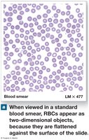
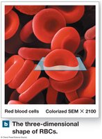
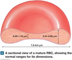
Biconcave discs – increase surface area for gas exchange.
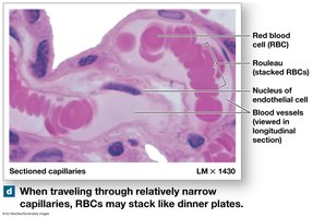
Form stacks (rouleaux) for smooth flow through capillaries.
Flexible to pass through small vessels.
Anucleate (no nucleus), lack mitochondria and ribosomes – cannot divide or repair.
Hemoglobin Structure and Function
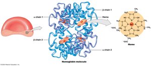
Composed of four polypeptide chains (2 alpha, 2 beta).
Each chain contains a heme group with an iron ion that binds oxygen.
Oxygen-bound form: Oxyhemoglobin; without oxygen: Deoxyhemoglobin.
Can also bind carbon dioxide (carbaminohemoglobin).
RBC Life Cycle and Turnover
Average lifespan: ~120 days.
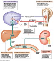
Destroyed by macrophages in spleen, liver, and bone marrow.
Hemoglobin is recycled: iron is stored or transported, heme is converted to biliverdin and then bilirubin (excreted in bile).
Disorders: Anemia (low hematocrit or hemoglobin), hemoglobinuria (hemoglobin in urine), hematuria (RBCs in urine).
Erythropoiesis (RBC Formation)
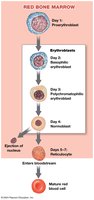
Occurs in red bone marrow (myeloid tissue) in adults.
Stem cells (hemocytoblasts) differentiate into myeloid and lymphoid stem cells.
Stages: Proerythroblast → Erythroblast → Normoblast (nucleus ejected) → Reticulocyte → Mature RBC.
Regulated by erythropoietin (EPO), secreted by kidneys and liver in response to hypoxia.
Requires amino acids, iron, and vitamins (especially folic acid).
Blood Types and Transfusion Compatibility
ABO Blood Group System
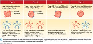
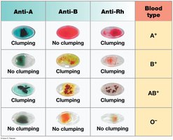
Determined by presence or absence of surface antigens (agglutinogens) A and B on RBCs.
Four main types: A, B, AB, O.
Plasma contains antibodies (agglutinins) against foreign antigens.
Rh Blood Group
Based on presence (Rh+) or absence (Rh−) of the D antigen.
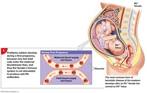
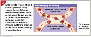
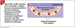
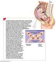
Rh− individuals can develop anti-Rh antibodies if exposed to Rh+ blood (sensitization).
Hemolytic disease of the newborn (HDN) can occur if an Rh− mother carries an Rh+ fetus.
Transfusion Reactions and Compatibility Testing
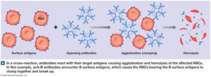
Cross-reaction: Occurs if donor and recipient blood types are incompatible, leading to agglutination and hemolysis.
Compatibility testing: Includes cross-match and blood typing for A, B, and Rh antigens.
Type O− is the universal donor; AB+ is the universal recipient.
White Blood Cells (Leukocytes)
Types and Functions
Defend against pathogens, remove toxins and wastes, attack abnormal cells.
Have nuclei and organelles, but lack hemoglobin.
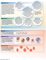
Most are found in connective tissues and lymphatic organs; only a small fraction circulates in blood (5,000–10,000/μL).
Classification of WBCs
Granular leukocytes: Neutrophils, eosinophils, basophils.
Agranular leukocytes: Monocytes, lymphocytes.
Neutrophils
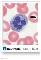
50–70% of WBCs; multilobed nucleus; pale granules.
First responders to injury; phagocytize bacteria; form pus.
Eosinophils
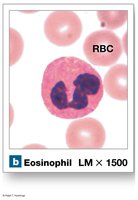
2–4% of WBCs; attack parasites; involved in allergic responses.
Release enzymes to reduce inflammation.
Basophils
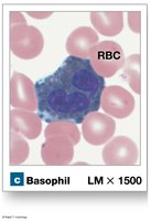
<1% of WBCs; release histamine (vasodilation) and heparin (anticoagulant).
Enhance inflammation at injury sites.
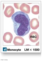
Monocytes
2–8% of WBCs; become macrophages in tissues; aggressive phagocytes.
Attract other immune cells and fibroblasts for tissue repair.
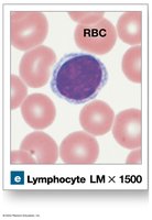
Lymphocytes
20–40% of WBCs; specific immunity; migrate between blood and tissues.
Types: T cells (cell-mediated immunity), B cells (antibody production), NK cells (immune surveillance).
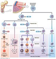
WBC Production and Regulation
Leukopoiesis: Formation of WBCs from hemocytoblasts in bone marrow.
Regulated by colony-stimulating factors (CSFs): Multi-CSF, GM-CSF, G-CSF, M-CSF.
Lymphocytes mature in lymphatic tissues (T cells in thymus, B and NK cells in bone marrow).
Platelets (Thrombocytes)
Structure, Function, and Production
Cell fragments involved in clotting; circulate 9–12 days; removed by spleen.
Normal count: 150,000–500,000/μL.
Functions: Release clotting chemicals, form temporary plugs, reduce vessel break size.
Produced by thrombocytopoiesis in bone marrow from megakaryocytes.
Regulated by thrombopoietin (TPO), interleukin-6 (IL-6), and Multi-CSF.
Hemostasis: Control of Blood Loss
Phases of Hemostasis
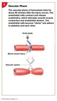
Vascular phase: Vascular spasm reduces blood flow; endothelial cells release factors for repair.
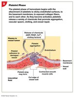
Platelet phase: Platelets adhere to exposed collagen, aggregate, and release chemicals to promote clotting.
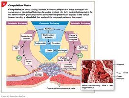
Coagulation phase: Cascade of clotting factors leads to fibrin formation and blood clot stabilization.
Coagulation Pathways
Extrinsic pathway: Triggered by tissue factor from damaged cells.
Intrinsic pathway: Initiated by exposure of blood to collagen.
Common pathway: Both pathways activate Factor X, leading to conversion of prothrombin to thrombin, and fibrinogen to fibrin.
Regulation and Disorders of Clotting
Anticoagulants (e.g., antithrombin III, heparin, thrombomodulin, prostacyclin) prevent excessive clotting.
Calcium ions and vitamin K are essential for clotting factor synthesis and function.
Disorders: Thrombocytopenia (low platelets), hemophilia (inherited bleeding), thrombophilia (excess clotting), deep vein thrombosis (DVT), pulmonary embolism.
Clot Retraction and Fibrinolysis
Clot retraction: Platelets contract to reduce the size of the clot and facilitate tissue repair.
Fibrinolysis: Plasminogen is converted to plasmin, which digests fibrin and dissolves the clot.
Additional info: These notes provide a comprehensive overview of blood structure, function, and clinical relevance, suitable for exam preparation in Anatomy & Physiology courses.
