 뒤로
뒤로Bones and Skeletal Tissues: Structure, Function, and Disorders
스터디 가이드 - 스마트 노트
 자료에 맞춘 맞춤형 노트, 핵심 정의, 예시, 맥락을 확장해 제공합니다.
Bones and Skeletal Tissues
Introduction
The skeletal system provides the framework for the human body, supporting movement, protecting organs, and serving as a reservoir for minerals. This chapter explores the structure, types, development, and disorders of bones and skeletal tissues.
Skeletal Cartilages
Basic Structure, Types, and Locations
Cartilage is a resilient, semi-rigid connective tissue found in various parts of the skeleton. It is avascular and receives nutrients by diffusion. There are three main types of cartilage:
Hyaline cartilage: Most abundant; provides support with flexibility and resilience. Found in articular surfaces, costal cartilages, respiratory structures, and nasal cartilage.
Elastic cartilage: Contains more elastic fibers, allowing flexibility. Found in the external ear and epiglottis.
Fibrocartilage: Highly compressible with great tensile strength. Found in intervertebral discs, pubic symphysis, and menisci of the knee.
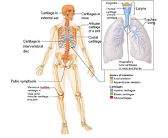
Locations: Cartilage is present in the external ear, nose, larynx, trachea, intervertebral discs, pubic symphysis, and articular surfaces of joints.
Growth of Cartilage
Appositional growth: Cells in the perichondrium secrete new matrix on the external surface of cartilage.
Interstitial growth: Chondrocytes within the cartilage divide and secrete new matrix, expanding the cartilage from within.
Cartilage growth typically ends during adolescence when the skeleton stops growing.
Functions of Bones
Major Functions
Support: Provides a framework for the body and cradles soft organs.
Protection: Protects vital organs (e.g., skull protects the brain, rib cage protects the thoracic organs).
Movement: Skeletal muscles attach to bones and use them as levers to produce movement.
Mineral storage: Reservoir for minerals, especially calcium and phosphate, which can be released into the bloodstream as needed.
Blood cell formation: Hematopoiesis occurs in red marrow cavities of certain bones.
Triglyceride (fat) storage: Fat is stored in yellow marrow cavities.
Hormone production: Osteocalcin, produced by bones, helps regulate bone formation and protects against obesity and diabetes.
Classification of Bones
Based on Shape
Long bones: Longer than they are wide (e.g., humerus, femur).
Short bones: Cube-shaped (e.g., carpals, tarsals).
Flat bones: Thin, flattened, and usually curved (e.g., sternum, ribs, skull bones).
Irregular bones: Complicated shapes (e.g., vertebrae, hip bones).
Bone Structure
Gross Anatomy
Bones are organs composed of bone tissue, nervous tissue, cartilage, fibrous connective tissue, muscle, and epithelial cells in blood vessels. There are two main types of bone tissue:
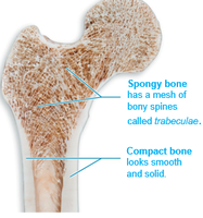
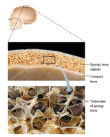
Compact bone: Dense outer layer that looks smooth and solid.
Spongy bone (trabecular bone): Internal layer with a honeycomb of small needle-like or flat pieces called trabeculae.
Structure of a Long Bone
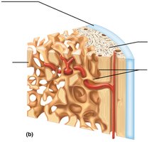
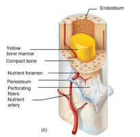
Diaphysis: Shaft; composed of compact bone surrounding a medullary cavity.
Epiphyses: Ends of the bone; contain spongy bone and are covered by articular cartilage.
Membranes: Periosteum (outer fibrous layer and inner osteogenic layer) and endosteum (lines internal bone surfaces).
Marrow: Yellow marrow (fat storage) in adults; red marrow (hematopoiesis) in children and some adult bones.
Bone Markings
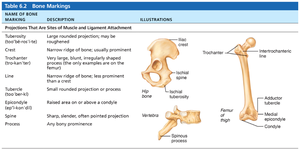
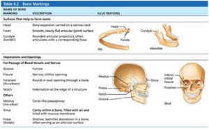
Bones display various markings that serve as sites of muscle, ligament, and tendon attachment, joint surfaces, or conduits for blood vessels and nerves.
Name of Bone Marking | Description | Illustrations |
|---|---|---|
Process | Any bony prominence | Spinous process |
Foramen | Round or oval opening through a bone | Skull foramen |
Fossa | Shallow, basinlike depression | Mandibular fossa |
Trochanter | Very large, blunt, irregularly shaped process | Femur of thigh |
Condyle | Rounded articular projection | Mandible condyle |
Microscopic Anatomy of Bone
Cells of Bone Tissue
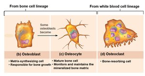
Bone contains five major cell types, all derived from the same basic cell lineage except osteoclasts:
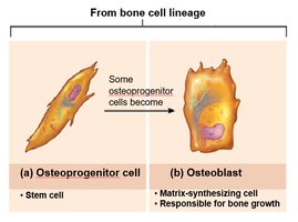
Osteoprogenitor cells: Stem cells that differentiate into osteoblasts.
Osteoblasts: Bone-forming cells that secrete bone matrix (osteoid).
Osteocytes: Mature bone cells that maintain the bone matrix.
Bone-lining cells: Flat cells on bone surfaces believed to help maintain the matrix.
Osteoclasts: Large, multinucleated cells that resorb (break down) bone.
Compact Bone Structure
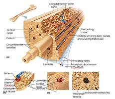
Osteon (Haversian system): Structural unit of compact bone; elongated cylinder parallel to the long axis of the bone.
Lamellae: Concentric rings of bone matrix.
Central (Haversian) canal: Contains blood vessels and nerves.
Perforating (Volkmann's) canals: Connect blood vessels and nerves of periosteum to those in the central canal.
Lacunae: Small cavities containing osteocytes.
Canaliculi: Tiny canals connecting lacunae, allowing for nutrient and waste exchange.
Spongy Bone Structure
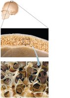
Spongy bone consists of trabeculae, which align along lines of stress to help resist bone stress. Spaces between trabeculae are filled with red or yellow marrow.
Chemical Composition of Bone
Organic and Inorganic Components
Organic components: Include cells (osteogenic cells, osteoblasts, osteocytes, osteoclasts) and osteoid (ground substance and collagen fibers), which provide flexibility and tensile strength.
Inorganic components: Mainly hydroxyapatites (mineral salts, primarily calcium phosphates), which provide hardness and resistance to compression.
Bone Development (Ossification)
Formation of the Bony Skeleton
Ossification (osteogenesis) is the process of bone tissue formation. In embryos, this leads to the formation of the bony skeleton; in adults, it serves mainly for bone remodeling and repair.
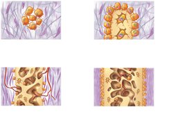
Endochondral ossification: Bone forms by replacing hyaline cartilage. Most bones below the base of the skull are formed this way.
Intramembranous ossification: Bone develops from a fibrous membrane. Forms flat bones such as the clavicles and cranial bones.
Postnatal Bone Growth
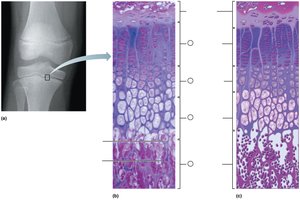
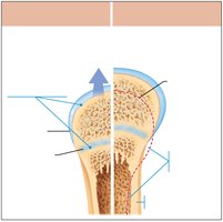
After birth, bones grow in length (interstitial growth) and thickness (appositional growth). Growth in length occurs at the epiphyseal plate through zones of proliferation, hypertrophy, calcification, and ossification.
Bone Remodeling
Bone Deposit and Resorption
Bone remodeling is a continuous process involving bone deposit by osteoblasts and bone resorption by osteoclasts. Remodeling helps maintain bone strength and mineral homeostasis.
Control of Remodeling
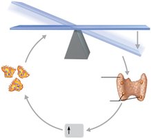
Hormonal control: Parathyroid hormone (PTH) increases blood calcium by stimulating osteoclasts to resorb bone. Calcitonin may play a minor role in lowering blood calcium.
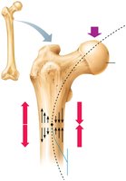
Mechanical stress: Bone adapts to the loads under which it is placed (Wolff's law).
Bone Repair
Fracture Classification
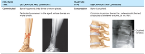
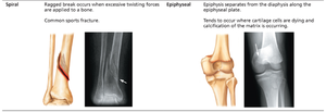
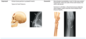
Fractures are classified by their position, completeness, orientation, and whether the bone ends penetrate the skin. Common types include comminuted, compression, spiral, epiphyseal, depressed, and greenstick fractures.
Fracture Type | Description and Comments |
|---|---|
Comminuted | Bone fragments into three or more pieces; common in aged bones. |
Compression | Bone is crushed; common in porous bones. |
Spiral | Ragged break due to excessive twisting forces; common sports fracture. |
Epiphyseal | Epiphysis separates from diaphysis along epiphyseal plate. |
Depressed | Broken bone portion is pressed inward; typical of skull fracture. |
Greenstick | Bone breaks incompletely; common in children. |
Fracture Treatment and Repair
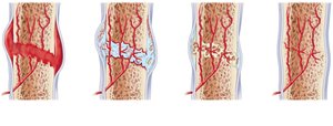
Bone repair involves four major stages:
Hematoma formation
Fibrocartilaginous (soft) callus formation
Bony callus formation
Bone remodeling
Bone Disorders
Osteomalacia and Rickets
Osteomalacia (in adults) and rickets (in children) are disorders caused by inadequate mineralization of bone matrix, often due to vitamin D or calcium deficiency. Bones become soft and weak.
Osteoporosis
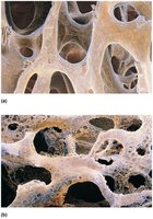
Osteoporosis is a condition in which bone resorption outpaces bone deposit, leading to porous and fragile bones. It is common in postmenopausal women and increases fracture risk.
Paget’s Disease
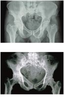
Paget’s disease is characterized by excessive and disorganized bone remodeling, resulting in structurally abnormal bone. The cause is unknown but may involve a viral trigger.
Additional info: This guide covers the essential concepts of bone and skeletal tissue structure, function, development, and common disorders, providing a foundation for further study in anatomy and physiology.
