 뒤로
뒤로Ch. 20 pt.2: Cardiac Physiology: Structure, Function, and Dynamics of the Heart
스터디 가이드 - 스마트 노트
 자료에 맞춘 맞춤형 노트, 핵심 정의, 예시, 맥락을 확장해 제공합니다.
Introduction to the Cardiovascular System
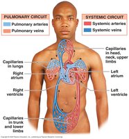
Overview of Circuits
The cardiovascular system is composed of two main circuits: the pulmonary and systemic circuits. These circuits are responsible for transporting blood throughout the body and facilitating gas and nutrient exchange.
Pulmonary Circuit: Carries blood to and from the lungs for gas exchange.
Systemic Circuit: Carries blood to and from the rest of the body.
Vessels:
Arteries: Efferent vessels that carry blood away from the heart.
Veins: Afferent vessels that carry blood to the heart.
Capillaries: Sites of gas and nutrient exchange.
Characteristics of Cardiac Cells
Nodal and Contractile Cells
Cardiac muscle tissue consists of two main cell populations: nodal cells and contractile cells.
Nodal Cells:
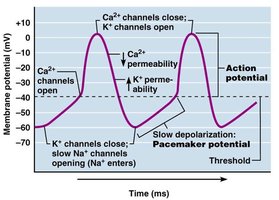
Pacemaker cells that spontaneously depolarize and generate action potentials without external influence.
Exhibit an unstable resting membrane potential.
Contractile Cells:
Have a stable resting membrane potential.
Intercalated disks allow cells to be chemically linked, facilitating coordinated contraction.
Cardiac Action Potentials
Nodal Cell Action Potentials
Nodal cells exhibit a "slow response" action potential characterized by a slow initial depolarization phase and repolarization to an unstable, slowly depolarizing "resting" potential. 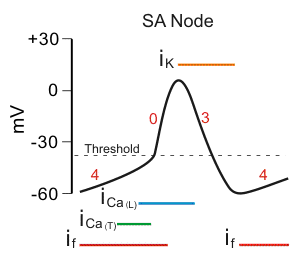
Contractile Cell Action Potentials
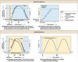
Contractile cells have a stable resting potential at -90mV and a plateau phase, resulting in a long absolute refractory period. 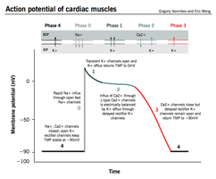
Cardiac Muscle Contraction vs Skeletal Muscle
Comparison and Role of Calcium
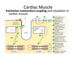
Cardiac muscle contraction is similar to skeletal muscle except for the role of calcium ions.
20% of calcium ions required for contraction enter during the plateau phase.
Arrival of extracellular Ca2+ triggers release of calcium from the sarcoplasmic reticulum (SR).
Cardiac muscle is highly sensitive to extracellular Ca2+ concentrations.
The Conducting System of the Heart
Components and Function
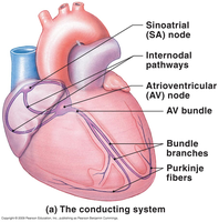
The conducting system consists of specialized cardiac muscle cells that initiate and distribute electrical impulses, stimulating contraction.
SA Node: Depolarizes first, establishing heart rate.
AV Node: Delays impulse, allowing atrial contraction.
AV Bundle, Bundle Branches, Purkinje Fibers: Distribute stimulus through myocardium.
Impulse Conduction
SA node initiates depolarization, spreading across both atria.
AV node depolarizes after a delay.
Impulse spreads through AV bundle, bundle branches, and Purkinje fibers to ventricles.
Abnormal Pacemaker Function
Arrhythmias and Ectopic Pacemakers
Arrhythmias: Abnormal rhythm or beat of the heart.
Bradycardia: Abnormally slow heart rate.
Tachycardia: Abnormally fast heart rate.
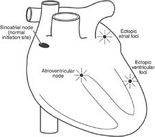
Ectopic Pacemaker: Abnormal cells generate high rates of action potentials, bypassing the conducting system and disrupting ventricular contractions.
The Electrocardiogram (ECG/EKG)
Recording and Interpretation
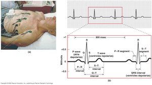
An ECG records electrical events in the heart, helping diagnose damage and arrhythmias.
P wave: Atrial depolarization.
QRS complex: Ventricular depolarization.
T wave: Ventricular repolarization.
P–R interval: Start of atrial depolarization to start of QRS complex.
Q–T interval: Ventricular depolarization to ventricular repolarization.
Energy for Cardiac Contractions
Aerobic Metabolism and Oxygen Supply
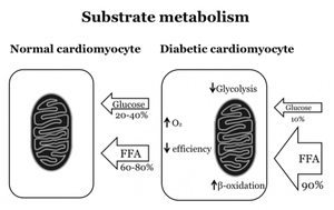
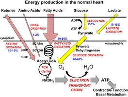
The heart requires large amounts of ATP, primarily produced through aerobic metabolism.
Energy is derived from mitochondrial breakdown of fatty acids and glucose.
Oxygen is supplied by circulating hemoglobin and stored in myoglobin within cardiac muscle.
Coronary Circulation and Heart Disease
Coronary Artery Disease (CAD)
Coronary circulation provides blood to heart musculature.
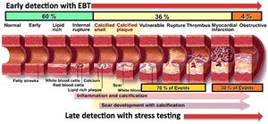
CAD involves partial or complete blockage of coronary arteries, leading to ischemia and reduced cardiac performance.
Insufficient oxygen delivery results in hypoxia and ischemia, potentially causing cell death.
The Cardiac Cycle
Phases and Pressure-Volume Relationships
The cardiac cycle is the period between the start of one heartbeat and the next, divided into systole and diastole.
Systole: Chamber contracts and pushes blood.
Diastole: Chamber relaxes and fills with blood.
Blood moves from areas of higher to lower pressure.
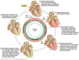
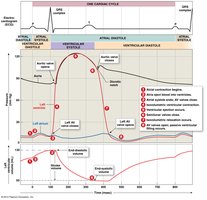
Phases of the Cardiac Cycle
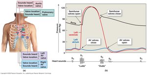
Atrial Systole: Atria contract, AV valves open, blood ejected into ventricles.
Atrial Diastole: Atria relax.
Ventricular Systole: Ventricles contract, AV valves close, semilunar valves open for ejection.
Ventricular Diastole: Ventricles relax, all valves closed, passive filling occurs.
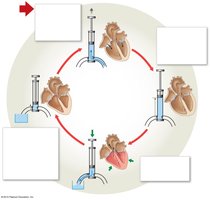
Pressure-Volume Loop
Graphical Representation
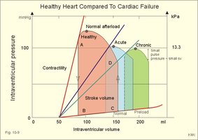
Pressure-volume loop graphs illustrate changes in pressure and volume during a single cardiac cycle and can show alterations due to disease.
Maximal pressure decreases with loss of contractile cells.
Volume shifts right as blood remains in ventricle due to decreased contractility.
Heart Sounds
Valvular Events
Lubb: Closing of AV valves.
Dubb: Closing of semilunar valves.
Gurgling sounds may indicate infection or heart murmur.
Cardiodynamics
Key Volumes and Formulas
Cardiodynamics refers to the movement and force generated by cardiac contractions.
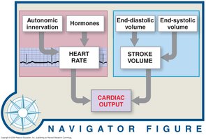
End-diastolic volume (EDV): Volume of blood in ventricle at end of diastole.
End-systolic volume (ESV): Volume remaining after systole.
Stroke volume (SV): Amount of blood ejected per beat.
Ejection fraction: Percentage of EDV represented by SV.
Cardiac Output
Amount of blood pumped by the left ventricle in one minute.
Formula:
Factors Affecting Heart Rate and Stroke Volume
Regulation of Heart Rate
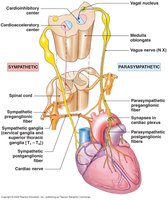
Parasympathetic: Acetylcholine (ACH) via vagus nerve decreases heart rate.
Sympathetic: Norepinephrine (NE) and epinephrine (EPI) increase heart rate.
Hormones: Thyroid hormones, NE, and EPI affect rate.
Venous Return: Bainbridge reflex increases heart rate with increased venous return.
Medulla: Cardioacceleratory and cardioinhibitory centers regulate autonomic output.
Factors Affecting Stroke Volume
Preload: Degree of stretch of cardiac muscle before contraction.
Contractility: Force of contraction at a given preload.
Afterload: Pressure the heart must overcome to eject blood.
Summary of Cardiac Physiology
Heart anatomy and electrophysiology
ECG interpretation
Cardiac action potentials
Conduction system
Excitation-contraction coupling
Cardiac cycle and pressure/volume loops
Regulation of cardiac output
Practice Questions
Cardiac output of the right side of the heart is what percentage of the cardiac output of the left side of the heart? Answer: 100% (Cardiac output of left and right sides should be equal)
From outermost to inner, name the 3 distinct layers of the heart.
Walk through blood flow as it would move through the heart if you start at the Vena Cava’s.
What are the key steps in an action potential of a SA nodal cell? Ventricle cell?
Additional info:
Academic context was added to clarify the role of calcium, the phases of the cardiac cycle, and the regulation of cardiac output.
