 뒤로
뒤로Cell Cycle and Mitosis: Structure, Phases, and Microscopic Identification
스터디 가이드 - 스마트 노트
 자료에 맞춘 맞춤형 노트, 핵심 정의, 예시, 맥락을 확장해 제공합니다.
Cell Cycle Overview
Definition and Importance
The cell cycle is the series of events that cells undergo from their formation to their division into two daughter cells. It is essential for growth, tissue repair, and reproduction in multicellular organisms. - Key Point 1: The cell cycle consists of two main phases: Interphase and the Mitotic (M) phase. - Key Point 2: Interphase is the period of cell growth and DNA replication, while the M phase includes mitosis and cytokinesis. - Example: The cell cycle ensures that each daughter cell receives a complete set of genetic material. 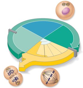
Interphase Subphases
Interphase is subdivided into three distinct subphases, each with specific cellular activities. - G1 (Gap 1): Vigorous cell growth and metabolism. - S (Synthesis): DNA replication occurs, preparing the cell for division. - G2 (Gap 2): Final preparations for cell division, including synthesis of proteins and organelles. 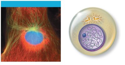
Mitosis: Nuclear Division
Definition and Purpose
Mitosis is the process of nuclear division in which replicated chromosomes are distributed to two daughter nuclei. It ensures genetic continuity and is followed by cytokinesis, the division of the cytoplasm. - Key Point 1: Mitosis consists of four main stages: Prophase, Metaphase, Anaphase, and Telophase. - Key Point 2: Each stage is characterized by specific structural changes in the chromosomes and cell. 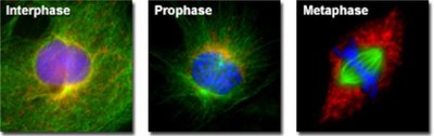
Prophase
Prophase is the first stage of mitosis, marked by the condensation of chromatin into visible chromosomes. - Early Prophase: Chromosomes become visible, each consisting of two sister chromatids joined at the centromere. Centrosomes migrate to opposite poles, and the mitotic spindle begins to form. - Late Prophase: The nuclear envelope fragments, and spindle fibers attach to the kinetochores of chromosomes. 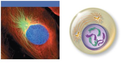
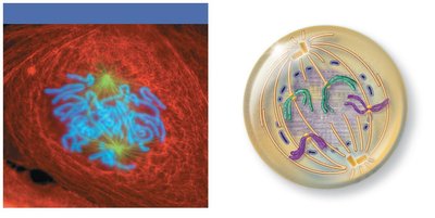
Metaphase
During metaphase, chromosomes align at the cell's equator, forming the metaphase plate. - Key Point: Spindle fibers ensure that each chromosome is attached and ready for separation. - Example: The metaphase plate is a critical checkpoint for proper chromosome segregation. 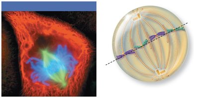
Anaphase
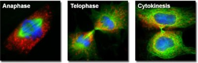
Anaphase is the shortest stage of mitosis, where sister chromatids are separated and pulled toward opposite poles. - Key Point: Motor proteins and spindle fibers facilitate the movement of chromosomes. - Example: Chromosomes appear V-shaped as they move apart. 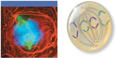
Telophase
Telophase marks the end of nuclear division, with chromosomes uncoiling and new nuclear envelopes forming. - Key Point: Nucleoli reappear, and the spindle apparatus disassembles. 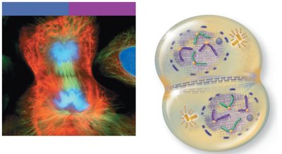
Cytokinesis
Cytokinesis is the division of the cytoplasm, resulting in two genetically identical daughter cells. - Key Point: A contractile ring of actin microfilaments forms a cleavage furrow, pinching the cell in two. 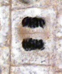
Microscopic Identification of Mitosis Stages
Allium Root Tip as a Model
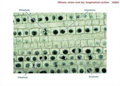
The Allium (onion) root tip is commonly used to study mitosis due to its high rate of cell division. - Key Point: Each stage of mitosis can be identified by distinct chromosomal arrangements under the microscope. 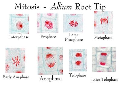
Longitudinal Section Analysis
Microscopic images of onion root tips show cells in various stages of the cell cycle. - Key Point: Interphase cells have a distinct nucleus, while mitotic cells show condensed chromosomes. - Example: Metaphase cells display chromosomes aligned at the center, anaphase cells show chromosomes moving apart, and telophase cells exhibit two nuclei. 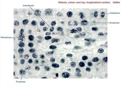
Summary Table: Mitosis Stages and Key Features
Stage | Main Features | Microscopic Appearance |
|---|---|---|
Interphase | Chromatin dispersed, nucleus intact | Large, round nucleus |
Prophase | Chromosomes condense, spindle forms | Chromosomes visible, nuclear envelope breaks |
Metaphase | Chromosomes align at equator | Chromosomes in a line at cell center |
Anaphase | Sister chromatids separate | Chromosomes move to poles |
Telophase | Nuclear envelope reforms, chromosomes decondense | Two nuclei, chromosomes less distinct |
Cytokinesis | Cytoplasm divides | Two separate cells |
Key Terms and Concepts
Chromatin: The uncondensed form of DNA found during interphase.
Chromosome: Condensed DNA structure visible during mitosis.
Centromere: Region where sister chromatids are joined.
Mitotic Spindle: Structure that separates chromosomes during mitosis.
Kinetochore: Protein structure on chromosomes where spindle fibers attach.
Cleavage Furrow: Indentation that begins cytokinesis.
Equations and Checkpoints
Cell Cycle Checkpoints
The cell cycle is regulated by checkpoints to ensure proper division. - G1 Checkpoint: Ensures cell is ready for DNA synthesis. - G2 Checkpoint: Ensures cell is ready for mitosis. - M Checkpoint: Ensures chromosomes are properly attached to spindle before separation.
DNA Replication Formula
The amount of DNA doubles during the S phase:
Applications and Clinical Relevance
Importance in Health and Disease
Proper regulation of the cell cycle and mitosis is crucial for normal development and prevention of diseases such as cancer. - Example: Uncontrolled cell division leads to tumor formation. Additional info: The cell cycle and mitosis are foundational concepts for understanding tissue growth, regeneration, and pathology in Anatomy & Physiology. ----------------------------------------
