 뒤로
뒤로Cell Structure and Function: Foundations of Anatomy & Physiology
스터디 가이드 - 스마트 노트
 자료에 맞춘 맞춤형 노트, 핵심 정의, 예시, 맥락을 확장해 제공합니다.
Cell Structure and Function
Introduction to Cell Theory
Cells are the fundamental units of life, forming the basis for all structure and function in living organisms. The cell theory provides the foundational principles for understanding biology and anatomy.
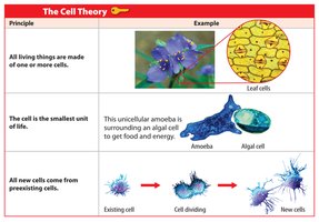
All living things are made of one or more cells. This principle highlights the universality of cells in all forms of life, from unicellular organisms to complex multicellular organisms.
The cell is the smallest unit of life. Cells carry out all necessary life processes, including metabolism, growth, and reproduction.
All new cells arise from preexisting cells. This principle explains cellular continuity through division and reproduction.
Microscopy and Cell Diversity
Techniques for Viewing Cells
Different microscopy techniques provide unique perspectives on cell structure and function:
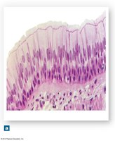
Light Microscopy (LM): Useful for viewing living cells and tissues at lower magnification.
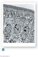
Transmission Electron Microscopy (TEM): Provides detailed images of internal cell structures and organelles at high magnification.
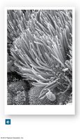
Scanning Electron Microscopy (SEM): Offers detailed views of cell and tissue surfaces.
Diversity of Cell Types
Cells in the human body exhibit a wide range of shapes and functions, reflecting their specialized roles. Examples include muscle cells, blood cells, neurons, fat cells, and reproductive cells (oocyte and sperm).
Somatic cells: All body cells except reproductive cells.
Sex cells: Oocytes (egg cells) and sperm cells, involved in reproduction.
Structure-function relationship: The shape and internal organization of a cell are closely related to its specific function.
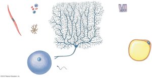
Cell Anatomy: Structure and Organelles
Overview of a Typical Cell
A typical eukaryotic cell contains various organelles, each with specialized functions essential for cell survival and activity.
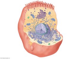
Summary Table: Anatomy of a Representative Cell
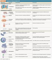
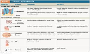
The following table summarizes the main structures, their composition, and functions in a typical cell:
Structure | Composition | Function(s) |
|---|---|---|
Plasmalemma (Cell Membrane) | Lipid bilayer with proteins, cholesterol, and carbohydrates | Isolation, protection, sensitivity, support, controls entry/exit of materials |
Cytosol | Fluid component of cytoplasm | Distributes materials by diffusion; site of chemical reactions |
Cytoskeleton | Proteins organized as filaments or slender tubes | Strength, support, movement of cellular structures and materials |
Microvilli | Membrane extensions containing microfilaments | Increase surface area for absorption |
Centrosome/Centrioles | Cytoplasm containing two centrioles at right angles | Essential for movement of chromosomes during cell division |
Cilia | Membrane extensions containing microtubules | Movement of materials over cell surface |
Ribosomes | RNA + proteins; fixed or free | Protein synthesis |
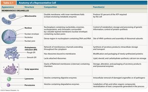
Mitochondria | Double membrane with inner folds (cristae) | Produce 95% of ATP required by the cell |
Nucleus | Nucleoplasm with DNA, nucleotides, enzymes, proteins | Control of metabolism, storage and processing of genetic information |
Endoplasmic Reticulum (ER) | Network of membranous channels | Synthesis of secretory products, intracellular storage and transport |
Golgi Apparatus | Stacks of flattened membranes (cisternae) | Storage, alteration, and packaging of secretory products and lysosomal enzymes |
Lysosomes | Vesicles containing digestive enzymes | Removal of damaged organelles or pathogens |
Peroxisomes | Vesicles containing degradative enzymes | Catabolism of fats and other organic compounds; neutralization of toxic compounds |
Plasmalemma (Cell Membrane) Structure
Phospholipid Bilayer and Membrane Proteins
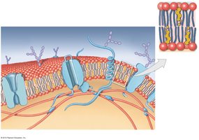
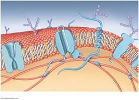
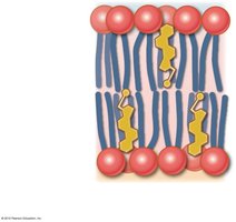
The cell membrane is primarily composed of a phospholipid bilayer with embedded proteins, cholesterol, and carbohydrates. This structure provides selective permeability and fluidity to the membrane.
Phospholipids: Form the basic structure, with hydrophilic heads facing outward and hydrophobic tails inward.
Cholesterol: Stabilizes membrane fluidity and integrity.
Integral and peripheral proteins: Serve as channels, receptors, and enzymes.
Glycocalyx: Carbohydrate-rich area involved in cell recognition and protection.
Membrane Transport Mechanisms
Passive and Active Processes
Substances move across the cell membrane by passive (no energy required) or active (energy required) processes.
Passive processes: Diffusion, osmosis, and facilitated diffusion.
Active processes: Active transport, endocytosis, and exocytosis.
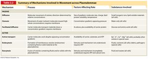
Mechanism | Process | Factors Affecting Rate | Substances Involved |
|---|---|---|---|
Diffusion | Movement of molecules from high to low concentration | Gradient size, membrane permeability, molecule size/charge, temperature | Gases, small inorganic ions, lipid-soluble materials |
Osmosis | Diffusion of water across a selectively permeable membrane | Solute concentration gradient, opposing pressure | Water |
Facilitated Diffusion | Passive transport via carrier proteins | Gradient size, carrier protein availability | Glucose, amino acids |
Active Transport | Carrier proteins move substances against gradient using ATP | Carrier protein and ATP availability | Na+, K+, Ca2+, Mg2+ |
Endocytosis | Vesicular transport into cell (phagocytosis, pinocytosis, receptor-mediated) | Stimulus, receptor presence | Fluids, macromolecules, pathogens |
Exocytosis | Vesicular transport out of cell | Stimulus, ATP, Ca2+ | Cellular wastes, secretory products |
Specialized Cell Structures
Cytoskeleton and Surface Extensions
The cytoskeleton provides structural support and facilitates movement within the cell. Surface extensions such as microvilli, cilia, and flagella serve specialized functions.
Microvilli: Increase surface area for absorption.
Cilia: Move fluids or materials across the cell surface.
Flagella: Propel sperm cells through fluid.
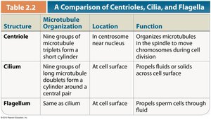
Structure | Microtubule Organization | Location | Function |
|---|---|---|---|
Centrioles | Nine groups of microtubule triplets form a short cylinder | In centrosome near nucleus | Organizes microtubules in the spindle to move chromosomes during cell division |
Cilia | Nine groups of microtubule doublets surround a central pair | At cell surface | Propels fluids or solids across cell surface |
Flagella | Same as cilium | At cell surface | Propels sperm cells through fluid |
Conclusion
Understanding cell structure and function is essential for the study of anatomy and physiology. The diversity of cell types, specialized organelles, and membrane transport mechanisms underlie the complexity and adaptability of living organisms.
