뒤로
뒤로Cell Structure and Function: Mini-Textbook Study Notes
스터디 가이드 - 스마트 노트
 자료에 맞춘 맞춤형 노트, 핵심 정의, 예시, 맥락을 확장해 제공합니다.
Cell Structure and Function
Introduction to Microscopes
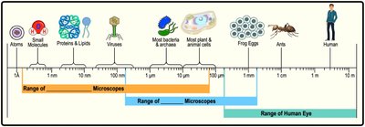
Microscopes are essential tools in biology, allowing visualization of objects too small for the human eye, such as cells and microbes. There are two main types of microscopes: light microscopes and electron microscopes.
Light Microscopes: Use visible light to magnify small objects, suitable for most plant and animal cells.
Electron Microscopes: Use electron beams for higher magnification, enabling visualization of structures like proteins, viruses, and cell organelles.
Range of Human Eye: Limited to objects larger than about 0.1 mm, such as frog eggs and ants.
Range of Microscopes: Light microscopes cover most cells; electron microscopes cover molecules and viruses.
Types of Electron Microscopes
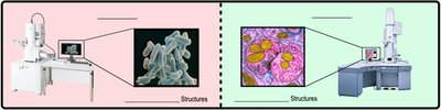
Scanning Electron Microscope (SEM): Visualizes external cell surfaces.
Transmission Electron Microscope (TEM): Visualizes internal cell structures.
Prokaryotic and Eukaryotic Cells
Domains of Life
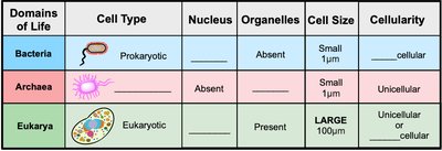
All living organisms are classified into three domains: Bacteria, Archaea, and Eukarya. These domains differ in cell type, presence of nucleus and organelles, cell size, and cellularity.
Domains of Life | Cell Type | Nucleus | Organelles | Cell Size | Cellularity |
|---|---|---|---|---|---|
Bacteria | Prokaryotic | Absent | Absent | Small (1 μm) | Unicellular |
Archaea | Prokaryotic | Absent | Absent | Small (1 μm) | Unicellular |
Eukarya | Eukaryotic | Present | Present | LARGE (100 μm) | Unicellular or Multicellular |
Features of Bacterial Cells
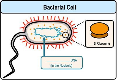
Bacteria are the most abundant and diverse organisms on Earth. Their DNA is circular and located in a region called the nucleoid. Bacteria possess small (70S) ribosomes and divide by binary fission.
Nucleoid: Region where bacterial DNA is found.
Ribosomes: Small, responsible for protein synthesis.
Features of Eukaryotic Cells
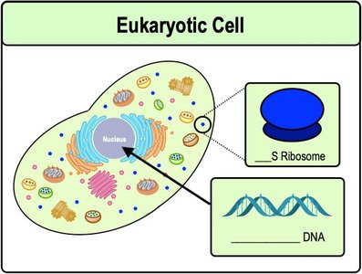
Eukaryotic cells contain a nucleus and several membrane-bound organelles. Their DNA is linear and located inside the nucleus. Eukaryotes have large (80S) ribosomes and divide by mitosis and cytokinesis.
Nucleus: Packages genetic material.
Ribosomes: Larger than prokaryotic ribosomes.
Prokaryotic vs. Eukaryotic Cells
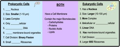
Prokaryotic and eukaryotic cells differ in complexity, size, and cellular structures. Both types share some features, such as a cell membrane and major biomolecules.
Prokaryotic Cells | BOTH | Eukaryotic Cells |
|---|---|---|
No nucleus | Cell membrane | Has nucleus |
Smaller (1-10 μm) | Carbohydrates, proteins, nucleic acids, lipids | Larger (10-100 μm) |
Less complex | More complex | |
Only unicellular | Unicellular or multicellular | |
Circular DNA | Linear DNA | |
No membrane-bound organelles | Has membrane-bound organelles | |
Binary fission | Mitosis | |
Small ribosomes | Larger 80S ribosomes |
Biological Membranes
Structure and Function
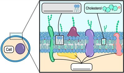
Biological membranes are primarily composed of amphipathic phospholipids, proteins, and cholesterol. The fluid mosaic model describes membranes as dynamic structures with proteins embedded in a phospholipid bilayer.
Phospholipid Bilayer: Provides structural foundation.
Proteins: Embedded and move laterally.
Cholesterol: Modulates membrane fluidity.
Types of Membrane Proteins
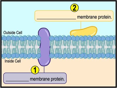
Membrane proteins are classified as integral (spanning the bilayer) or peripheral (attached to the membrane surface).
Integral Membrane Proteins: Span the entire bilayer.
Peripheral Membrane Proteins: Located on the perimeter.
Functions of Membrane Proteins
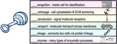
Recognition: Marks cell for identification.
Anchorage: Anchors cytoskeleton and extracellular matrix.
Transduction: Signal molecule receptors.
Transport: Molecular transport across membrane.
Linkage: Connects cells via protein linkage.
Enzymes: Catalyze enzymatic processes.
Concentration Gradients and Diffusion
Concentration Gradients
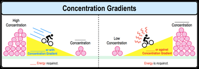
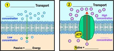
A concentration gradient is the difference in concentration of a substance between two areas. Molecules move down their gradient (from high to low concentration) passively, or up their gradient (from low to high concentration) actively, requiring energy.
Diffusion
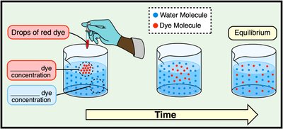
Diffusion is the movement of a substance from an area of high concentration to an area of low concentration, driven by the natural tendency of molecules to reach equilibrium.
Passive Process: No energy required.
Equilibrium: Achieved when concentrations are equal.
Membrane Transport
Selectively Permeable Membranes
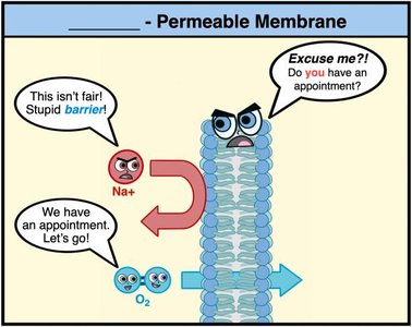
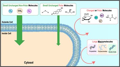
Biological membranes are selectively permeable, allowing certain molecules to cross while blocking others. Small, uncharged, nonpolar molecules can freely diffuse; large, charged, or polar molecules require protein facilitation.

Types of Membrane Transport
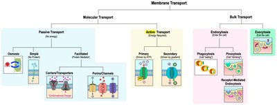
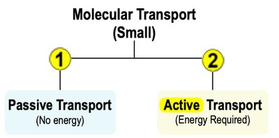
Membrane transport is categorized as passive (no energy) or active (requires energy). Passive transport moves molecules down their concentration gradient, while active transport moves molecules against their gradient.
Classes of Membrane Transport Proteins
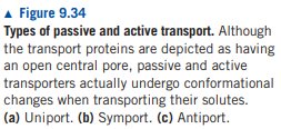
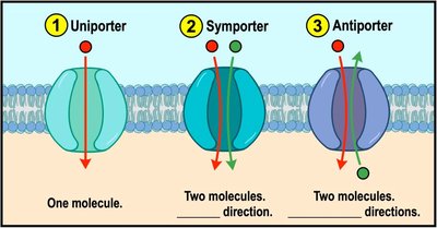
Uniporters: Transport one molecule at a time in one direction.
Symporters: Cotransport two or more molecules in the same direction.
Antiporters: Cotransport two or more molecules in opposite directions.
Osmosis and Tonicity
Osmosis
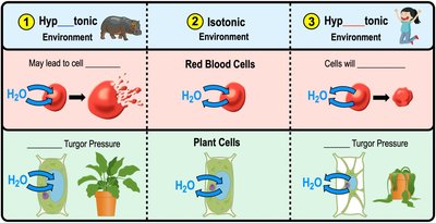
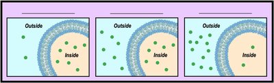
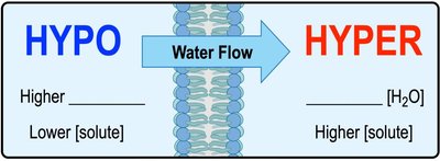
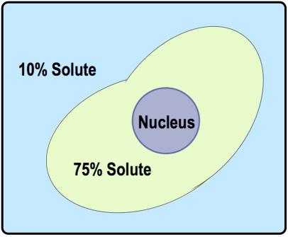
Osmosis is the passive diffusion of water across a semi-permeable membrane. The direction of water flow depends on the relative concentration of solutes in the solutions.
Hypotonic: Lower solute concentration outside the cell.
Isotonic: Equal solute concentrations inside and outside.
Hypertonic: Higher solute concentration outside the cell.
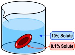
Direction of Osmosis
Water moves from hypotonic to hypertonic solutions if solutes cannot diffuse across the membrane. Water always moves from higher to lower water concentration.
Environmental Tonicity Effects
Hypotonic Environments: Water enters cells, causing swelling and possible lysis; preferred by plant cells for turgor pressure.
Isotonic Environments: Water enters and exits at equal rates; preferred by animal cells.
Hypertonic Environments: Water exits cells, causing dehydration.