뒤로
뒤로Cells: The Living Units – Structure and Function of Cytoplasmic Organelles
스터디 가이드 - 스마트 노트
 자료에 맞춘 맞춤형 노트, 핵심 정의, 예시, 맥락을 확장해 제공합니다.
Cells: The Living Units
Cytoplasm
The cytoplasm is the cellular material located between the plasma membrane and the nucleus. It is the site of most cellular activities and consists of three main components:
Cytosol: A viscous, semitransparent fluid composed mainly of water, proteins, salts, sugars, and other solutes.
Organelles: Specialized structures that perform specific metabolic functions within the cell. Organelles can be membranous (e.g., mitochondria, endoplasmic reticulum) or nonmembranous (e.g., ribosomes, centrioles).
Inclusions: Chemical substances that may or may not be present, depending on cell type (e.g., glycogen granules, lipid droplets, pigments).
Cytoplasmic Organelles
Membranous Organelles
Mitochondria
Peroxisomes
Lysosomes
Endoplasmic Reticulum (ER)
Golgi Apparatus
Nonmembranous Organelles
Cytoskeleton
Centrioles
Ribosomes
Membranes allow for compartmentalization, which is crucial for the specialization of cellular functions.
Mitochondria
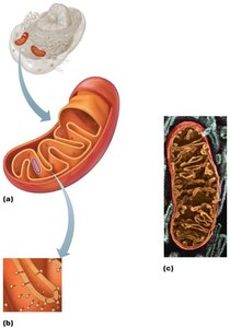
Mitochondria are double-membrane organelles with inner folds called cristae. They are the primary site of ATP production via aerobic cellular respiration, a process that requires oxygen. Mitochondria contain their own DNA, RNA, and ribosomes, and can replicate independently through a process called fission. Their structure and function are similar to bacteria, supporting the endosymbiotic theory.
Ribosomes
Ribosomes are small granules composed of protein and ribosomal RNA (rRNA). They are the sites of protein synthesis and exist in two forms:
Free ribosomes: Synthesize proteins that function within the cytosol or other organelles.
Membrane-bound ribosomes: Attached to the rough ER; synthesize proteins destined for membranes, lysosomes, or export from the cell.
Endoplasmic Reticulum (ER)
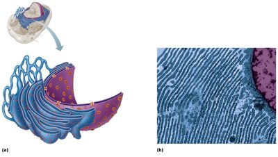
The ER is a network of interconnected tubes and parallel membranes enclosing fluid-filled cavities called cisterns. It is continuous with the outer nuclear membrane and exists in two forms:
Rough ER: Studded with ribosomes; synthesizes all secreted proteins, membrane proteins, and phospholipids. Newly synthesized proteins are enclosed in vesicles and sent to the Golgi apparatus.
Smooth ER: Lacks ribosomes; involved in lipid metabolism, synthesis of steroid hormones, detoxification of drugs and chemicals, glycogen breakdown, and calcium storage.
Golgi Apparatus
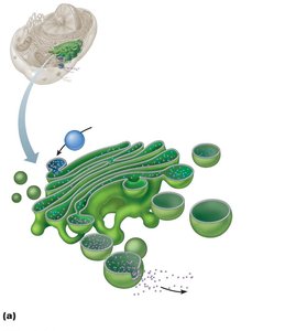
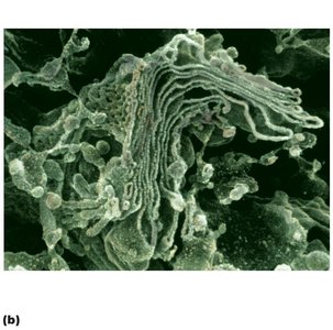
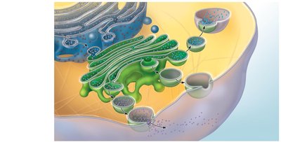
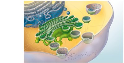
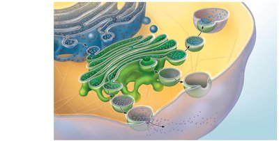
The Golgi apparatus consists of stacked, flattened membranous sacs. It modifies, concentrates, and packages proteins and lipids received from the rough ER. Transport vesicles from the ER fuse with the cis face (receiving side) of the Golgi, and proteins are processed and sorted for delivery. Three types of vesicles bud from the trans face (shipping side):
Secretory vesicles: Release export proteins by exocytosis.
Vesicles for membranes: Deliver lipids and transmembrane proteins to the plasma membrane or organelles.
Lysosomes: Contain digestive enzymes and remain in the cell.
Protein Synthesis and Trafficking
Proteins synthesized on the rough ER are transported to the Golgi apparatus, where they are modified and sorted for their final destinations. The sequence of events includes:
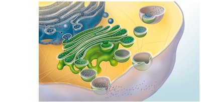
Vesicles containing proteins pinch off from the rough ER and fuse with the Golgi apparatus.
Proteins are modified within the Golgi compartments.
Proteins are packaged into different vesicle types for secretion, incorporation into membranes, or delivery to lysosomes.
Peroxisomes
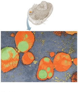
Peroxisomes are membranous sacs containing oxidases and catalases. They detoxify harmful substances, catalyze and synthesize fatty acids, and neutralize free radicals. Oxidases convert substances to hydrogen peroxide (H2O2), which is then converted to water and oxygen by catalases.
Lysosomes
Lysosomes are spherical membranous organelles containing digestive enzymes (acid hydrolases). They serve as safe sites for intracellular digestion, breaking down ingested bacteria, viruses, toxins, nonfunctional organelles, and cellular debris. Lysosomes also play a role in autolysis (self-destruction of cells) and bone resorption to release calcium.
Endomembrane System
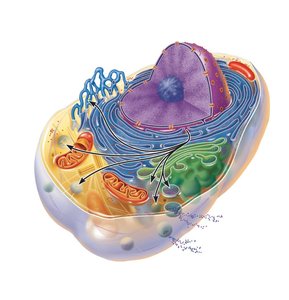
The endomembrane system is a network of organelles that work together to produce, degrade, store, and export biological molecules, as well as degrade harmful substances. It includes the ER, Golgi apparatus, secretory vesicles, lysosomes, and the nuclear and plasma membranes.
Cytoskeleton
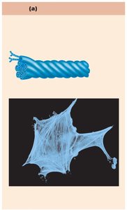
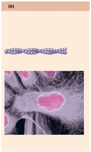
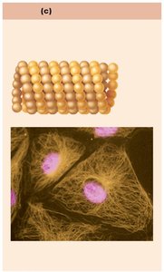
The cytoskeleton is an elaborate network of protein filaments that extends throughout the cytosol, providing structural support, shape, and movement for the cell. It consists of three main types of filaments:
Microfilaments: Thinnest elements, composed of actin; involved in cell movement, shape changes, and endocytosis/exocytosis.
Intermediate filaments: Tough, ropelike fibers; provide tensile strength and resist pulling forces; attach to desmosomes.
Microtubules: Largest elements, hollow tubes made of tubulin; determine cell shape, organelle distribution, and serve as tracks for motor proteins.
Motor Proteins
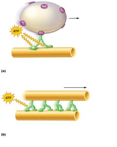
Motor proteins are complexes that move organelles and vesicles along cytoskeletal tracks, powered by ATP. They are essential for intracellular transport and cell motility, including muscle contraction and cilia movement.
Centrosome and Centrioles
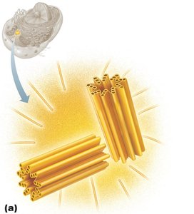
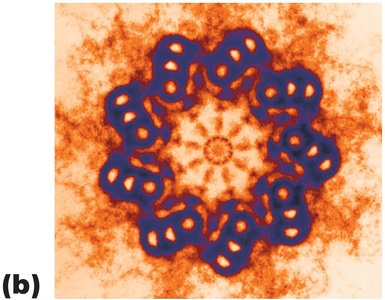
The centrosome is the cell center near the nucleus, responsible for organizing microtubules and the mitotic spindle during cell division. It contains paired centrioles, which are barrel-shaped structures made of microtubules. Centrioles also form the bases of cilia and flagella.
Cilia and Flagella
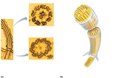
Cilia and flagella are motile cellular extensions containing microtubules and motor proteins. Cilia move substances across cell surfaces, while flagella (e.g., sperm tail) propel entire cells. The base of each is formed by a basal body derived from centrioles. Cilia exhibit a coordinated power and recovery stroke to move materials.
Microvilli
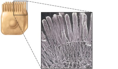
Microvilli are minute, fingerlike extensions of the plasma membrane that increase surface area for absorption. They contain a core of actin filaments for structural support.
Nucleus
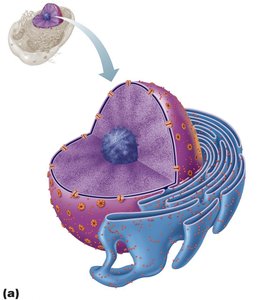
The nucleus is the largest organelle and serves as the genetic library of the cell, containing DNA that codes for nearly all cellular proteins. It responds to signals and dictates the types and amounts of proteins synthesized. Most cells are uninucleate, but some are multinucleate (e.g., skeletal muscle), and mature red blood cells are anucleate.
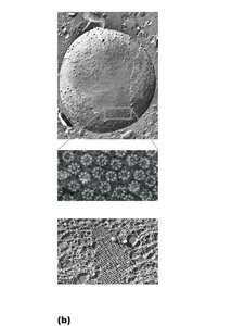
Nuclear envelope: Double-membrane barrier with pores for molecular transport; outer membrane is continuous with rough ER.
Nucleoli: Sites of rRNA synthesis and ribosome assembly; usually one or two per cell.
Chromatin: Threadlike DNA-protein complex; condenses into chromosomes during cell division.
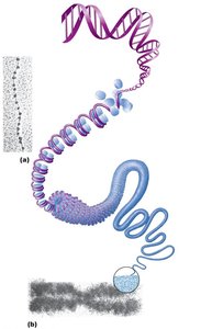
Chromatin Structure
Chromatin consists of DNA (30%), histone proteins (60%), and RNA (10%). It is organized into nucleosomes, which are DNA wrapped around histone proteins. Chromatin condenses into chromosomes during cell division.