 뒤로
뒤로Chapter 1: An Introduction to Anatomy and Physiology - Study Notes
스터디 가이드 - 스마트 노트
 자료에 맞춘 맞춤형 노트, 핵심 정의, 예시, 맥락을 확장해 제공합니다.
Introduction to Anatomy & Physiology
Overview and Learning Outcomes
This chapter introduces the foundational concepts of anatomy and physiology, emphasizing their interrelationship and the importance of mastering terminology and organizational levels. Students will learn to use textbook resources, define key terms, and understand the structure and function of the human body.
Anatomy: Study of internal and external body structures and their physical relationships.
Physiology: Study of how living organisms perform vital functions.
Principle of Complementarity: Structure and function are closely related; specific functions are performed by specific structures.
Anatomy and Physiology Specialties
Types of Anatomy
Anatomy is divided into gross (macroscopic) and microscopic anatomy, each with distinct areas of focus.
Gross Anatomy: Examines large structures visible to the naked eye.
Regional Anatomy: Focuses on specific body areas.
Systemic Anatomy: Studies organ systems.
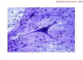
Microscopic Anatomy: Requires magnification to study cells and tissues.
Cytology: Study of cell structure.
Histology: Study of tissue structure.
Types of Physiology
Cell Physiology: Function of cells and their chemical processes.
Organ Physiology: Function of specific organs.
Systemic Physiology: Function of organ systems.
Pathological Physiology: Effects of diseases on organs or systems.
Levels of Organization in the Human Body
Hierarchical Structure
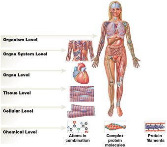
The human body is organized into six major levels, from the simplest chemical level to the most complex organism level.
Chemical Level: Atoms and molecules.
Cellular Level: Cells, the smallest living units.
Tissue Level: Groups of cells performing specific functions. Four types: epithelial, connective, muscle, neural.
Organ Level: Two or more tissues working together (e.g., heart).
Organ System Level: Groups of organs interacting for a particular function (e.g., cardiovascular system).
Organism Level: Individual life form.


Anatomical Terminology
Standard Anatomical Position and Body Regions
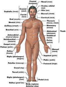
Understanding anatomical terminology is essential for describing locations, positions, and relationships of body parts.
Anatomical Position: Standing upright, hands at sides, palms forward, feet together.
Supine: Lying face up.
Prone: Lying face down.
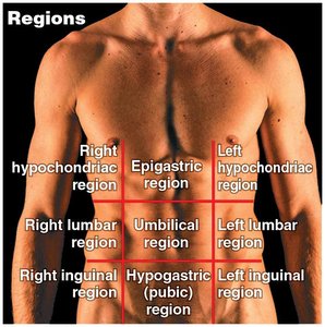
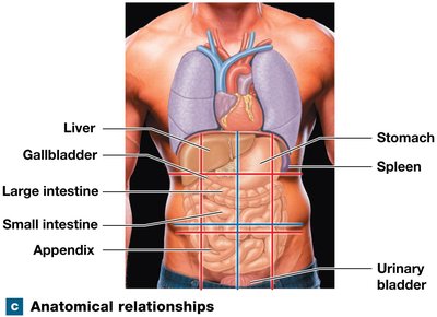
Abdominopelvic Quadrants and Regions
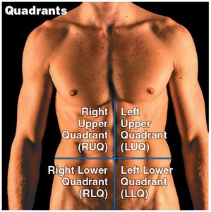
The abdominopelvic area is divided for clinical and anatomical purposes.
Quadrants: Right Upper (RUQ), Left Upper (LUQ), Right Lower (RLQ), Left Lower (LLQ).
Regions: Epigastric, Right/Left hypochondriac, Umbilical, Right/Left lumbar, Hypogastric, Right/Left inguinal.
Directional Terms
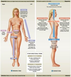
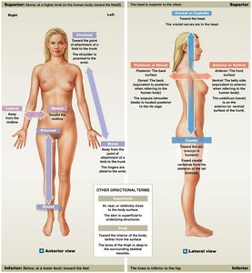
Directional terms describe the relative location of body regions and structures.
Superior: Toward the head.
Inferior: Toward the lower part or away from the head.
Anterior (Ventral): Nearer to the front.
Posterior (Dorsal): Nearer to the back.
Medial: Nearer to the midline.
Lateral: Toward the side, farther from the midline.
Ipsilateral: Same side.
Contralateral: Opposite sides.
Proximal: Nearer to the point of attachment.
Distal: Farther from the point of attachment.
Superficial: Toward or on the surface.
Deep: Away from the surface.
Sectional Anatomy and Medical Imaging
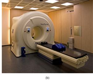
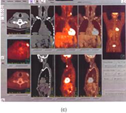
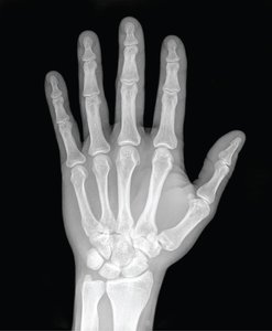
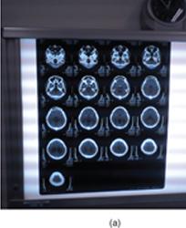
Sectional anatomy involves slicing through three-dimensional objects to visualize internal organization, important for interpreting medical scans.
MRI (Magnetic Resonance Imaging): Noninvasive, uses magnetic fields and radio waves, no radiation exposure.
PET (Positron Emission Tomography): Uses radioactive substances, diagnoses various conditions.
X-ray: High-energy electromagnetic radiation, penetrates solids.
CT (Computed Tomography): Analyzes cross-sectional X-rays, higher radiation dose than X-rays.
Sectional Planes
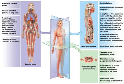
Sectional planes are used to describe slices through the body.
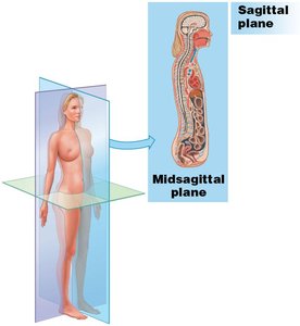
Frontal (Coronal) Plane: Divides body into anterior and posterior portions.
Sagittal Plane: Divides body into left and right portions.
Midsagittal: Lies in the middle.
Parasagittal: Offset from the middle.
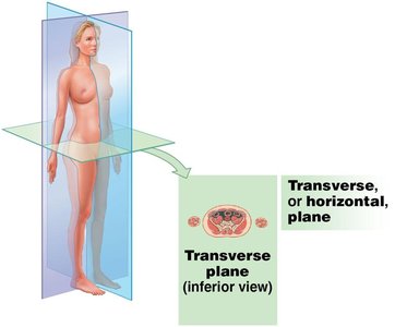
Transverse (Horizontal) Plane: Divides body into superior and inferior portions.
Body Cavities
Major Body Cavities and Their Functions
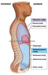
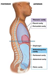
Body cavities are closed, fluid-filled spaces lined by serous membranes, containing vital organs (viscera) and serving protective and functional roles.
Functions: Protect organs from shocks, permit changes in size and shape.
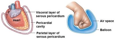
Serous Membrane (Serosa): Parietal layer lines cavity, visceral layer covers organ, serous fluid reduces friction.
Thoracic and Abdominopelvic Cavities
Thoracic Cavity: Divided from abdominopelvic cavity by diaphragm. Contains pleural cavities (lungs), pericardial cavity (heart), and mediastinum (connective tissue stabilizing organs).
Abdominopelvic Cavity: Extends from diaphragm to pelvis. Contains abdominal cavity, pelvic cavity, and peritoneal cavity (lined with peritoneum).
Retroperitoneal Space: Area between parietal peritoneum and back of body wall; contains pancreas and kidneys.
Pelvic Cavity: Contains reproductive organs, rectum, urinary bladder.
Homeostasis
Concept and Mechanisms
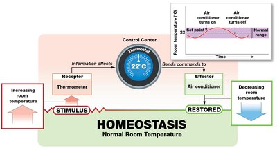
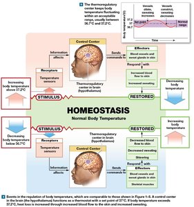
Homeostasis refers to the continuous physiological processes that maintain a stable internal environment. Systems respond to changes to keep variables within normal ranges.
Receptor: Sensor detecting stimulus.
Control Center: Processes information and sends commands.
Effector: Cell or organ carrying out commands.
Set Point: Desired value for internal conditions.
Negative and Positive Feedback
Feedback mechanisms regulate homeostasis by opposing or enhancing changes from normal.
Negative Feedback: Opposes variation from normal; effector response negates original stimulus, maintaining variables within normal range.
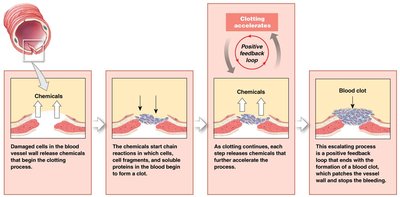
Positive Feedback: Enhances variation from normal; response amplifies original change, used for rapid completion of processes (e.g., blood clotting).
Summary Table: Levels of Organization
Level | Description | Example |
|---|---|---|
Chemical | Atoms and molecules | Protein molecules |
Cellular | Smallest living units | Muscle cell |
Tissue | Group of cells | Muscle tissue |
Organ | Two or more tissues | Heart |
Organ System | Group of organs | Cardiovascular system |
Organism | Individual life form | Human |
Key Terms and Definitions
Anatomy: Study of structure.
Physiology: Study of function.
Homeostasis: Maintenance of stable internal environment.
Serous Membrane: Lines cavities, covers organs.
Feedback: Regulatory mechanism.
