 뒤로
뒤로Chapter 1: An Introduction to Anatomy and Physiology – Structured Study Notes
스터디 가이드 - 스마트 노트
 자료에 맞춘 맞춤형 노트, 핵심 정의, 예시, 맥락을 확장해 제공합니다.
Chapter 1: An Introduction to Anatomy and Physiology
Why Study Anatomy and Physiology?
Anatomy and physiology are foundational sciences for all health professions and biology-based careers. Understanding these subjects enables individuals to make informed health choices, interpret medical news, and become more knowledgeable about their own bodies.
Anatomy: The study of internal and external body structures and their physical relationships among other body parts.
Physiology: The study of how living organisms perform their vital functions.
Integration: Structure and function are closely related; anatomical features often determine physiological capabilities.
Applications: Useful for careers in medicine, allied health, research, and personal health management.
Fields of Anatomy and Physiology
Anatomy and physiology are divided into several subfields based on the scale of observation and function.
Human Anatomy: Study of the structures of the human body.
Gross Anatomy (Macroscopic Anatomy): Examination of large structures visible without a microscope (e.g., cadaver dissection).
Microscopic Anatomy: Examination of structures only visible with a microscope (e.g., histology – study of tissues).
Human Physiology: Study of the functions of the human body.
Levels of Organization in the Human Body
The human body is organized into hierarchical levels, each with increasing complexity.
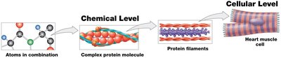
Chemical Level
The most basic level, consisting of atoms and molecules. Atoms are the smallest stable units of matter, and molecules are combinations of atoms (e.g., H2O).
Atoms: Fundamental units of matter.
Molecules: Two or more atoms bonded together.
Cellular Level
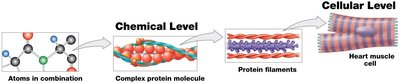
Cells are the smallest living units in the human body. Humans are eukaryotes, meaning their cells contain organelles.
Cells: Basic units of life; perform essential functions.
Eukaryotic Cells: Contain membrane-bound organelles.
Tissue Level
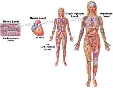
A tissue is a group of cells working together to perform one or more specific functions. Tissues often consist of related cell types.
Tissue: Group of similar cells performing a function.
Example: Cardiac muscle tissue in the heart.
Organ Level
An organ is a distinct structure composed of two or more tissue types that performs specific functions.
Organ: Structure with specialized functions (e.g., heart).
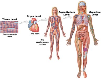
Organ System Level
An organ system is a group of organs that interact to perform specific functions.
Organ System: Group of organs working together (e.g., cardiovascular system).
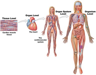
Organism Level
The organism level represents the individual living life form, such as a human.
Organism: Complete living entity.
Example: Human being.
Organ Systems of the Human Body
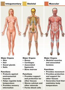
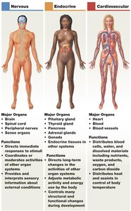
The human body contains multiple organ systems, each with distinct functions and major organs.
Integumentary System: Skin, hair, nails; protects against environmental hazards.
Skeletal System: Bones, cartilages, ligaments; provides support and protection.
Muscular System: Skeletal muscles; enables movement and generates heat.
Nervous System: Brain, spinal cord, nerves; directs immediate responses to stimuli.
Endocrine System: Glands; regulates metabolic activity and development.
Cardiovascular System: Heart, blood vessels; distributes blood and regulates temperature.
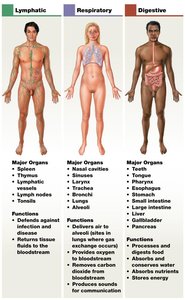
Lymphatic System: Spleen, lymph nodes; defends against infection.
Respiratory System: Lungs, airways; delivers air and removes carbon dioxide.
Digestive System: Stomach, intestines; processes food and absorbs nutrients.
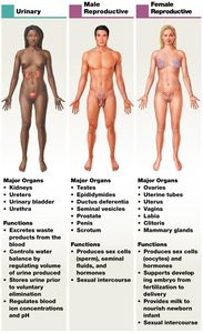
Urinary System: Kidneys, bladder; eliminates waste and regulates water balance.
Reproductive System: Ovaries/testes; produces sex cells and hormones.
Anatomical Terminology
Using standardized anatomical terms eliminates ambiguity and ensures clear communication in healthcare and research.
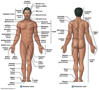
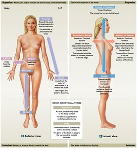
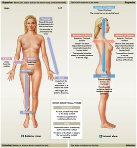
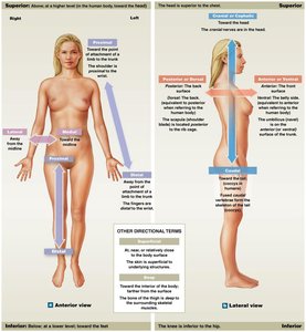
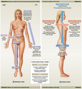
Anatomical Position: Standard reference position; body upright, feet together, hands at sides, palms forward.
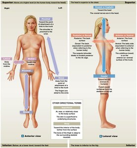
Anterior (ventral): Toward the front of the body.
Posterior (dorsal): Toward the back of the body.
Superior (cranial): Above or higher than another structure; toward the head.
Inferior (caudal): Below or lower than another structure; toward the feet.
Lateral: Away from the midline.
Medial: Toward the midline.
Proximal: Toward the point of attachment.
Distal: Away from the point of attachment.
Superficial: Near the surface.
Deep: Farther from the surface.
Prone: Face-down orientation.
Supine: Face-up orientation.
Sectional Anatomy
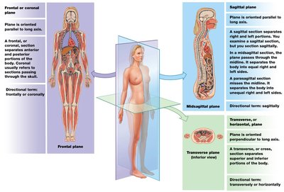
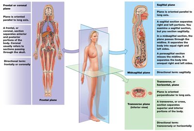
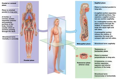
Sectional anatomy involves viewing the body along specific planes to understand internal structures. Medical imaging relies on these planes for accurate interpretation.
Section: Single view or slice along a plane.
Sectional Plane: Imaginary 2-D surface passing through the body.
Frontal (Coronal) Plane: Divides body into anterior and posterior portions.
Sagittal Plane: Divides body into right and left portions; midsagittal is exactly in the middle, parasagittal is offset.
Transverse Plane: Divides body into superior and inferior portions.
Body Cavities
Body cavities are closed, fluid-filled spaces lined by serous membranes. They protect organs and allow for changes in organ size and shape.
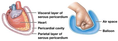
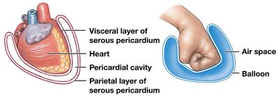
Serous Membranes (Serosa): Line cavity walls and cover organ surfaces; consist of visceral and parietal layers.
Visceral Layer: Directly covers the organ.
Parietal Layer: Lines the inner surface of the cavity wall.
Serous Fluid: Moistens layers and reduces friction.
Thoracic Cavity
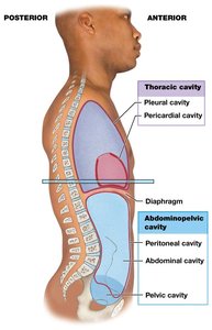
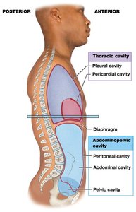
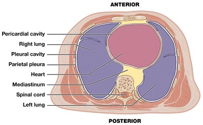
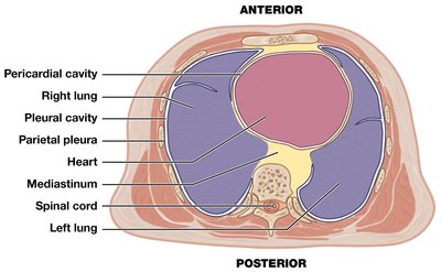
The thoracic cavity is deep to the chest wall and separated from the abdominopelvic cavity by the diaphragm. It contains pleural cavities (lungs), pericardial cavity (heart), and the mediastinum (connective tissue supporting major structures).
Pleura: Serous membrane lining pleural cavities; visceral pleura covers lungs, parietal pleura lines body wall.
Serous Pericardium: Lines pericardial cavity; visceral layer covers heart, parietal layer lines mediastinum.
Abdominopelvic Cavity
The abdominopelvic cavity is deep to the abdominal and pelvic walls, separated from the thoracic cavity by the diaphragm. It is subdivided into the abdominal cavity (liver, stomach, spleen, intestines) and pelvic cavity (bladder, reproductive organs).
Peritoneum: Serous membrane lining the peritoneal cavity; parietal peritoneum lines body wall, visceral peritoneum covers digestive organs.
Retroperitoneal Organs: Located between peritoneal lining and muscular wall (e.g., kidneys, pancreas).
Abdominopelvic Quadrants and Regions
Clinicians use quadrants and regions to describe locations of pain, injuries, or abnormalities.
Quadrants: Four areas for clinical reference.
Regions: More precise anatomical divisions used in research.
Homeostasis
Homeostasis is the continuous physiological process that maintains a stable internal environment. It is essential for survival and involves regulatory mechanisms that respond to changes in internal and external conditions.
Homeostatic Regulation: Adjustment of physiological systems to maintain homeostasis.
Three Main Parts: Receptor (sensor), Control Center (processes information), Effector (responds to commands).
Set Point: Desired value range for physiological conditions (e.g., body temperature).
Negative Feedback
Negative feedback mechanisms keep physiological conditions within their set points by opposing deviations from normal values.
Response: Effectors counteract changes, restoring normal range.
Example: Thermoregulation – body temperature is regulated by negative feedback.
Positive Feedback
Positive feedback mechanisms enhance or intensify the original stimulus, often used when a rapid response is needed to restore homeostasis.
Response: Effectors amplify changes until a definitive endpoint is reached.
Examples: Blood clotting and labor/delivery.
Summary Table: Levels of Organization
Level | Description | Example |
|---|---|---|
Chemical | Atoms and molecules | H2O, proteins |
Cellular | Smallest living units | Heart muscle cell |
Tissue | Group of similar cells | Cardiac muscle tissue |
Organ | Structure with specialized function | Heart |
Organ System | Group of organs working together | Cardiovascular system |
Organism | Complete living entity | Human |
Summary Table: Homeostatic Regulation
Component | Function |
|---|---|
Receptor | Detects stimulus or change |
Control Center | Processes information and sends commands |
Effector | Responds to commands, opposes or enhances stimulus |
Summary Table: Feedback Mechanisms
Type | Response | Example |
|---|---|---|
Negative Feedback | Opposes deviation from set point | Thermoregulation |
Positive Feedback | Enhances deviation until endpoint | Blood clotting, labor |
Additional info: These notes provide a comprehensive overview of Chapter 1, suitable for exam preparation and foundational understanding in Anatomy & Physiology.
