 뒤로
뒤로Chapter 1: An Introduction to Anatomy and Physiology – Study Notes
스터디 가이드 - 스마트 노트
 자료에 맞춘 맞춤형 노트, 핵심 정의, 예시, 맥락을 확장해 제공합니다.
Introduction to Anatomy and Physiology
This chapter introduces the foundational concepts of anatomy and physiology, focusing on the characteristics of living organisms, the relationship between structure and function, and the organization of the human body. Understanding these basics is essential for further study in the field of health sciences.
Basic Functions of Living Organisms
Common Functions
Responsiveness: The ability to detect and respond to changes in the environment (also called irritability). Example: withdrawing a hand from a hot surface. Adaptability refers to long-term adjustments, such as growing a thicker coat in winter.
Growth: Increase in size due to cell growth or addition of new cells. Cellular differentiation is the process by which cells become specialized for specific functions.
Reproduction: Production of new generations of the same organism.
Movement: Can be internal (e.g., transporting blood) or external (e.g., walking).
Metabolism: The sum of all chemical reactions in the body, providing energy for other functions. Includes processes such as respiration, digestion, and excretion.
Metabolic Processes
Respiration: Absorption, transport, and use of oxygen by cells.
Digestion: Breakdown of complex foods into simpler compounds for absorption.
Excretion: Elimination of waste products generated by metabolism.
Relationship Between Anatomy and Physiology
Definitions and Specialties
Anatomy: The study of internal and external structures and their physical relationships. Divided into:
Gross (Macroscopic) Anatomy: Structures visible to the naked eye (e.g., organs, tissues).
Microscopic Anatomy: Structures requiring magnification (e.g., cells, tissues).
Physiology: The study of function in living organisms. Includes specialties such as cell physiology, special physiology (specific organs), systemic physiology (organ systems), and pathological physiology (effects of disease).
Key Point: Anatomy and physiology are closely related; structure determines function, and function is explained by structure.
Levels of Organization in the Human Body
The human body is organized into hierarchical levels, each increasing in complexity.
Chemical Level: Atoms combine to form molecules. Molecular shape determines function.
Cellular Level: Cells are the smallest living units. Molecules interact to form cell structures.
Tissue Level: Groups of similar cells working together to perform specific functions.
Organ Level: Two or more tissues working together to perform specific functions.
Organ System Level: Organs interacting to perform complex functions.
Organism Level: All organ systems working together to maintain life and health.









The 11 Organ Systems of the Human Body
The human body is composed of 11 organ systems, each with specific functions essential for survival.
Organ System | Main Functions |
|---|---|
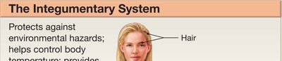
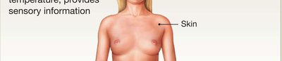
Integumentary | Protection, temperature regulation, sensory information |
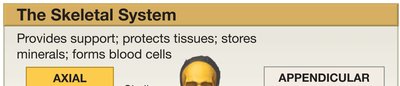
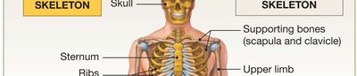
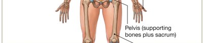
Skeletal | Support, protection, mineral storage, blood cell formation |
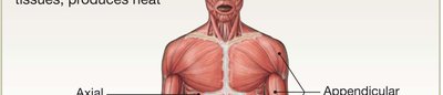
Muscular | Movement, protection, support, heat production |
Nervous | Immediate response to stimuli, coordination of organ systems, sensory information |
Endocrine | Long-term changes in organ systems, metabolic activity regulation |
Cardiovascular | Transport of cells and dissolved materials (nutrients, gases, wastes) |
Lymphatic | Defense against infection, returns tissue fluids to bloodstream |
Respiratory | Gas exchange, sound production |
Digestive | Digestion, absorption of nutrients, water absorption, waste elimination |
Urinary | Elimination of wastes, water balance, pH regulation |
Reproductive | Production of sex cells and hormones |




Homeostasis
Concept and Importance
Homeostasis: The maintenance of a stable internal environment. Essential for survival; its breakdown leads to illness or disease.
Maintained by the coordinated actions of cells, tissues, organs, and organ systems.
Homeostatic Regulation
Involves three main components:
Receptor: Detects changes (stimuli).
Control Center: Processes information and sends commands.
Effector: Responds to commands to oppose or enhance the stimulus.
Example: A thermostat system regulates room temperature using negative feedback.
Negative Feedback
Most common mechanism for maintaining homeostasis.
Response opposes the original stimulus, returning the system to normal.
Example: Thermoregulation (body temperature control).
Positive Feedback
Response reinforces the original stimulus, leading to an escalating cycle.
Occurs in processes that must be rapidly completed, such as blood clotting and childbirth.
Anatomical Terminology
Purpose and Use
Standardized language for describing body regions, positions, and directions.
Based on Latin and Greek roots.
Anatomical Position and Landmarks
Anatomical Position: Standing, hands at sides, palms forward, feet together.
Supine: Lying face up; Prone: Lying face down.
Abdominopelvic Quadrants and Regions
Quadrants: Four regions used clinically to locate pain or injury.
Regions: Nine regions used for more precise anatomical study.
Directional Terms
Describe the location of structures relative to anatomical position (e.g., anterior/ventral, posterior/dorsal).
Left and right refer to the subject's sides, not the observer's.
Sectional Anatomy
Helps understand three-dimensional structure using planes:
Frontal (Coronal) Plane: Divides body into anterior and posterior parts.
Sagittal Plane: Divides body into left and right parts (midsagittal = equal halves).
Transverse Plane: Divides body into superior and inferior parts.
Body Cavities of the Trunk
Major Body Cavities
Thoracic Cavity: Contains the heart (pericardial cavity) and lungs (pleural cavities).
Abdominopelvic Cavity: Contains digestive, urinary, and reproductive organs. Subdivided into abdominal and pelvic cavities.
Separated by the diaphragm.
Serous Membranes
Line body cavities and produce fluid to reduce friction.
Parietal Layer: Lines cavity wall; Visceral Layer: Covers organs.
Potential spaces exist between these layers.
Special Terms
Retroperitoneal: Organs located between the peritoneal lining and the dorsal body wall (e.g., kidneys).
Summary Table: Major Body Cavities and Their Contents
Cavity | Main Contents | Serous Membrane |
|---|---|---|
Pericardial | Heart | Pericardium |
Pleural (2) | Lungs | Pleura |
Abdominal | Digestive organs, spleen, kidneys (retroperitoneal) | Peritoneum |
Pelvic | Urinary bladder, reproductive organs, rectum | Peritoneum (partially) |
Example: An incision just inferior to the diaphragm opens the abdominopelvic cavity.
